CATEGORIES:
BiologyChemistryConstructionCultureEcologyEconomyElectronicsFinanceGeographyHistoryInformaticsLawMathematicsMechanicsMedicineOtherPedagogyPhilosophyPhysicsPolicyPsychologySociologySportTourism
The radio diagnostics of inflammatory diseases of the musculoskeletal system
Section contents:
Radiological signs of inflammatory damages of the musculoskeletal system: tuberculosis of bones and joints, osteomielitis, reumatoid arthritis, syphilitic defeat of bones.
The inflammatory diseases of the bones: tuberculosis of bones (tubercular osteitis) and joints (primary-osteal and primary-synovial form)
The tubercular osteitis
Radiological features: focuses of destruction (size 1.2-2.5 sm) located in epymetaphysis with irregular margins, the spongy sequestrum in a center of destruction maybe are located (as "piece of melting sugar"), osteoporosis (local, regionar), periostitis is absence, in child's age atrophy of bone is potential.
The joint tuberculosis accounts for 20% of all tubercular damages of bones.
The primary bone form runs in three phases(see fig12.1) : 1) a prearthritical phase corresponds to tubercular ostitis; 2) arthritical phase - a process passes from bone tissue to soft tissue and cartilaginous elements of joint, spreads quickly, causes the acute exudative reaction and excrescence of granulation tissue, destruction of joint cartilages and joint surfaces of epiphysises. After some expansion there is narrowing of roentgenologic joint space and regionarniy osteoporosis, a destructive process causes considerable deformations of joint surfaces of bone, resulting in their congruent lost, and in more widespread destructions come destructive dislocations; 3)postarthritical phase is completed by fibrotic ankylosis.
1 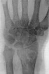 2
2  3
3 
Fig.12.1 1 - tubercular ostitis; 2 – arthritical phase; 3 - posatarthritical phase (fibrotic ankylosis)
The roentgenologic picture of the primary synovial form (see fig 12.2) in early stages corresponds to the picture of hydropsy of joint (expansion of joint space, regionary osteoporosis of bones, that form a joint, and thickening of joint capsule). Afterwards in 1-2 months in the places of attachment of capsule the small regional defects of irregular, rounded or oval form become noticeable. A thin cortical layer disappears and trabecules of the spongy substance resolve partly or fully. Destruction of cartilages is represented on pictures by narrowing of x-ray joint space. The stages of development of bone tuberculosis accoding to CORNEV P. G.: 1st- beginning, 2nd- height, 3rd- subsiding of process.
1 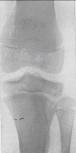 2
2 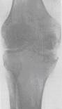
Fig.12.2 the primary-synovial form of joints tuberculosis: 1 – hydropsy of joints; 2 – destruction of joint cartilages and joint ends of bones.
The roentgenologic signs of tubercular spondilitis:
1) the decline of height of the inter-vertebral disk
2) the decline of height of the vertebral body (sign of pathological compression)
3) the destruction of the vertebral body in the type of separate focus, more frequent in the type of defect of edge in combination with destruction of the disk
4) the deformation of column with formation of a hump – angular kyphosis
5) the shades of abscess are observed in 80-90% cases in the defeat of thoracic department of column.
1 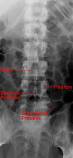 2
2 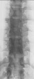 3
3 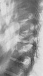
Fig.12.3. Lumbar department of column: 1 – norm; 2, 3 – tubercular spondilitis.
The tubercular coxitis (see fig 12.4). It the primary-bone disease usually - a primary tubercular focus arises up more frequent (64%) in bones of acetabulum. As a result of fracture of process in a joint there is destruction of joint cartilages, synovial sitll, joint bag, afterwards a process passes to other bone. Possible dystensional luxations in acute processes, as a result of distension of the joint bag by a plenty of exudate, at considerable destructions of bones`s joint ends there are destructive dislocations. In favourable course and timely medical treatment the process can end by some deformation of joint surfaces with saving of joint function; in the expressed destructions of joint surfaces a process ends with intracoxigeal pseudoartrosis or fibrotic ankylosis or destructive dislocation. X-ray signs: regionarniy osteoporosis, compression of joint bag, focus of destruction in bones of a joint.
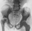
Fig.12.4. Tubercular coxitis
The Spina ventosa tuberculosa is the tuberculosis of diaphysis of tubular bones. The roentgenologic signs are deformation (thickening), destruction, sequestrum as a “piece of melting sugar”, periosteitis. The completions are disappearance of destruction and periosteitis.
Osteomyelitis (see fig 12.5) is the festering disease of bones, usually of staphylococcal etiology. We distinguish acute itmatogenic, gunshot, primary-chronic and secondary-chronic osteomielitis. Frequency of defeat: Thighbone - 46%, a shinbone is a 42% humeral bone - 10%, other bones - 3%.
The ordinary form of osteomielitis runs across in 4 phases: 1st- the phase of acute bone brain inflammation is a phlegmon, 2nd- fracture of process under the periosteum and formation of abscess; 3rd- necrosis of bone; 4th- phase of sequestration and reparation. X-ray signs appear on the 12-16 day of the disease: the focus of destruction in metadiaphysis is surrounded by osteosclerosis, in a center the sequestrum (corticalniy, central, penetrable, total), along a bone linear, fimbriated periosteitis. Gunshot osteomielitis is a result of development of festering infection in the gunshot fracture.
1  2
2  3
3 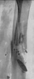
Fig.12.5 1 – types of sequestrum; 2 – acute osteomielitis; 3 – chronic osteomielitis; 4 –gunshot osteomielitis
The second-chronic osteomielitis is the result of acute hematogenic osteomielitis, is characterized by expressed bone-formation as the spacious areas of osteosclerosis, surrounded by the areas of liquid or normal bone tissue, sequestrum located in cavities with sclerostic contours, periostitis (fimbriated, crista-like, striped).
The atypical forms. Epiphysar osteomielitis meets in 72% cases under age of 2 years and is caused by strepto-, staphylo - and pneumococci. A process is more frequent by is localized in epiphysis thighs, runs acutely. Epiphysis collapses fully; there is suppuration of joint, rupture of abscess with formation of fistula. On a sciagram a thigh-bone is deformed, epiphysis and the part of metaphysis are absent; trochanter cavity of the bone is smoothed, femoral bone is in a state of dislocation up and is joined with an illum. The trochanter cavity in a process is not pulled in.
The chronic Garre’s primary-sclerotic osteomielitis (see fig 12.6): beginning is primary-chronic, the edema in connective tissues (infiltration of muscles) appears, limitation of mobility in surrounding joints. Roentgenologically: osteosclerosis, eburneation of (destructive foci, cavities, sequestrum are not present) and hyperostosis.
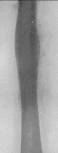
Fig.12.6 The Gare`s primary sclerotic osteomielitis
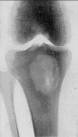
Fig.12.7 The Brodi`s abscess
The Brodi`s abscess (see fig 12.7): metaphysis large tubular bones (fibula - 80%) are struck in children or youth age. Roentgenologic: cavity by a diameter 2-3 sm with a clear contour is often surrounded by osteosclerosis; a sequestrum and periostitis are absent.
The reumatiod arthritis (see fig 12.8) will mainly strike the shallow joints of hands and feet. Roentgenologic: osteoporosis, bulge of separate trabecules, bulge of joint bag, narrowing of joint spaces. On the lateral edges of bones surfases there are regional excrescences. In the epiphysiar ends of bones there are cysts brightness. On edges of epiphysis there are bone excrescences as acute edges. Sometimes we can see squeezing in the center of joint cavity in which a joint head enters.
Fig.12.8 Reumatoid arthritis
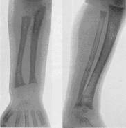
Fig.12.9 The syphilitic osteohondritis

Fig.12.10 Gummas syphilis
The syphilitic diseases of the bones( see fig 12.9,12.10). In early born syphilis there are the following forms:
1. Osteohondritis - this systemic syphilitic mage of to the skeleton in the areas of enchondral ossification. Roentgenologically 3 stages of changes are distinguished: 1st stage is expansion of area of previous calcification; 2nd - light stripe between the lines of area of calcification and bone (development of granulation tissue); 3rd - the expressed destructive changes, near the bone toothed, laceration, possible pathological fractures.
2. Periostitis is the periostal changes (stratification) will be disposed unevenly in relation to the sides of bone: on concave sides there are more powerful shades of stratifications. Osificational periostitis (plural, symmetrically located; often the systemic that the typical display of early born syphilis).
3. The gummas defects can be congenital and acquired. Roentgenologic are revealed out in the periostis or endostis destructive changes as oval or rounded foci of clearing up.
4. Falangitis. Defect of short ubular bones, which appear in the type of ostitis, osteomyelitis, periostitis, and osteohondritis. Roentgenologic more frequent are determined periostal stratification and sclerosis of the bone structure.
The defect of bones in late born syphilis is observed in 1-2 and even 4-5 year age and in more late age. There is in the type of plural periostitis and ostitis. There is deformation of bones.
The osteoartropathy at colagenosis will strike the symmetric, mainly shallow joints of hands and feet, there is their deformation and muscular atrophy.
The osteoartropathy at colagenosis has three phases.
1) The phase of the synoviitis: expansions of x-ray joint space, subdislocations or dislocations, increase of volume of connective tissues. Exudate in a joint can be defined earlier by means of the US and MRI visualisation, than on a sciagram.
2) The osteolytic phase: resolution of joint ends with gradual obliteration of marrowy channel, possible forming of neoarthrosis.
3) The osteoblastic phase: acutely expressed bone osteofites on the rest of incongrouent joint ends of bones.
The aseptic arthroso-arthritis arise in the metabolic disturbance (at a gout), exogenous and endogenous intoxications, hemorrhages in a joint (at haemophilia).
The roentgenologic sings are deformation of joints, decline of x-ray joint space, soubhondral sclerosis, and regional bone excrescences. In gout there are plural 3-5 mm in diameter bone defects (tofouses). The severe forms of deforming arthrosis are formed in course of time.
Questions for self-control:
1. Roentgenologic semiotics of the bones and joints diseases. 2. Osteomyelitis roentgenologic diagnostics|. Roentgenologic diagnostics of the osteomyelitis primary chronic| forms|shapes||. 3. Roentgenologic diagnostics of the bones and joints tuberculosis. Tubercular spondilitis| its|its| X-ray diagnostics|.
Section 13
Date: 2014-12-28; view: 1960
| <== previous page | | | next page ==> |
| The radio anatomy of the musculoskeletal system | | | The radio diagnostics of the musculosceletal system’s tumours |