CATEGORIES:
BiologyChemistryConstructionCultureEcologyEconomyElectronicsFinanceGeographyHistoryInformaticsLawMathematicsMechanicsMedicineOtherPedagogyPhilosophyPhysicsPolicyPsychologySociologySportTourism
The radio anatomy of the musculoskeletal system
The roentgenologic and CT visualisation allows getting information about bones and joints. It is explained by that bones are the richest depot in the organism of mineral salts (45% their composition - mineral, from them 85% - salts of calcium and phosphorus, 10% - potassium and carbon, 5% - magnesium). Mineral composition to increase the x-rays absorption. Adjoining soft tissues very poorly absorb the X-ray beam and make an natural backdrop for bone structure.
The sciagrams allows to show intravital image of bone. Roentgen-anatomy is the special branch of medical science, based on thorough knowledge of x-ray image, normal, topographical and pathological anatomy.
1  2
2  3
3 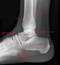
Fig.11.5 Normal bones: 1 – teenager; 2, 3 – adult.
The image of bones, joints and soft tissues in an axial projection is the feature of computer tomograms.
The skeleton of the grown up is composed of more than 200 bones ( see fig 11.6) - long, short, flat and aeriferous (air containing). Long bones are characterized by predominance of length over two other sizes, consist from diaphysis (middle part) and two epiphyses (joint extremities) - proximal and distal. Part of diaphysis which adjoins to epiphysis is called metaphysis. In the children between epiphysis and metaphysis the epiphyseal cartilage is located – on the sciagrams epiphyseal cartilage visualize as radiolucent line. The bone processes located on metaphysis and those having their own centers of ossification and tendons (of muscle) insertion site are named apophysis.
Tubular bones are short and long.
There is the spongy and compact substance of bone. The compact substance is on the all departments of bones, except for joint surfaces and is called a cortical (its most thickness in the middle of diaphysis, where the compact substance has the osteonic structure).
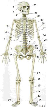
Fig.11.6. Skeletal System
1-Frontal bone;2-Parietal bone;3-Temporal bone;4-Maxilla;5-True rib;6-False rib;7-Vertebral column;8-Symphysis pubis;9-Pubic bone;10-Tarsal bones;11-Phalanges;12-Metatarsal bones;13-Talus;14-Fibula;15-Tibia;16-Patella;17-Femur;18-Ischium;19-Coccyx;20-Ilium;21-Sacrum;22-Carpal bones;23-Phalanges;24-Metacarpal bones;25-Ulna;26-Radius;27-Floating rib;28-Humerus;29-Sternum;30-Clavicle;31-Mandible;32Orbit
A bone grows longways due to an epiphyseal cartilage and breadthways due to the cambial layer of periosteum.
The union of bones. Structure of joints. There are immobile, a low-mobile and nonmobile connection of bones.
Three types of immobile connections differ by the type of connecting tissue: syndesmosis (bones of skull), synhondrosis (handle and body of breastbone and etc.) and synostosis.
The mobile connections of bones form joints. Their elements are: the joined surfaces of bones and joint cartilages, joint cavity, joint capsule and joint ligaments.
The sciagrams of bones and joints of children are characterized by the presence of:
- epiphysiar ossification center;
- radiolucent line of epiphyseal cartilage;
- wide joint space.
Plan of study of bones and joints sciagrams:
1. To estimate the position, form and size of bones;
2. To learn the contours of external and internal surfaces of cortical layer along bone;
3. To learn the bone structure;
4. To learn the condition and location of ossific nucleus, condition of bone grow plate (at children);
5. To learn correlation of joint ends of bones, size and form of joint space;
6. To learn a volume and structure of soft tissues around of bone.
The chart of sciagrams description
1. The method, visualisation area, projections.
2. The positions, sizes, form of bones.
3. The contours of external and internal surface of cortical layer.
4. The bone structure.
5. The reaction of periosteum.
6. The ossific nucleus and bone growth plate (at children).
7. The joint ends, form and sizes of joint space.
8. The soft tissues which surround a bone (joint).
9. The clinical-roentgenologic comment.
Symptons of bone pathology
| Symptoms of bone pathology, that are accompanied with reduction of bone substances in unit of volume | Symptoms of bone pathology, that are accompanied by the increase of bone substance in the unit of volume |
| Osteoporosis | Osteosclerosis |
| Atrophy | Hyperostosis, paraostosis |
| Destruction, sequestration | Periostitis |
| Expanded | Blastomatous growth |
Osteoporosis - is a reduction the bone substances in a unit of volume without the change bone sizes. Opposite to osteoporosis is osteosclerosis - this increase of bone substance in a unit of volume without the change bone sizes.
1 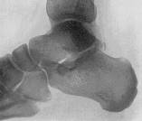 2
2  3
3  4
4 
Fig.11.7 Sciagrams of bones: 1 – normal structure of bones; 2 - osteoporosis; 3 – chart of osteoporosis; 4 – osteosclerosis, hyperostosis.
Radiological signs of osteoporosis and osteosclerosis:
| OSTEOPOROSIS | OSTEOSCLEROSIS |
| Decrease of bone density without reduction of the bone diameter, a bone becomes more radiolucent | Increase of bone density without reduction of the bone diameter, the spongy structure of bone disappears, a bone becoms more radiopaque |
| Thinning trabecules of bone and reduction of their quantity in a unit of bone volume | Thickening trabecules of bone and increase of their quantity in a unit of bone volume |
| Thinning of compact layer | Thickening of compact layer |
| Widening of marrowy channel | Narrowing of bones medullar channel |
Osteosclerosis and complete disappearance of bones medullar channel is named bone eburnation.
Osteoporosis by spreading may be: local, regional, widespread and systemic
Osteoporosis can be physiologic in old age, from inactivity of extremity, due to acute inflammatory processes and other. In acute processes it often is spotty, and in chronic -diffuse, widespread.
Osteosclerosis more frequent develops around an inflammatory process, metastases, after a trauma and other. Osteoporosis by spreading may be - local, limited, widespread and system.
| ATROPHY | HYPEROSTOSIS |
| Decrease of bone density with reduction of the bone diameter. | Increase of bone density with the increase of the bone diameter. |
| Thinning of compact layer with decrease of marrowy channel (concentric atrophy) and thinning of compact layer with increase of marrowy channel – excentric atrophy. Local atrophy is due to local pressure. | Thickening of bone due to periostal bone formation – ossification periostitis which coalesces with a compact layer. |
| Is accompanied with osteoporosis | Is accompanied with osteosclerosis |
The cause of the atrophy (see fig 11.8) is disturbance of trophism, intoxication, the inactivity and other causes that affected during to a long time.
1 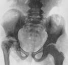 2
2 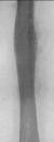
Fig.11.8 1 – atrophy of right hip and right half of pelvis; 2 – hyperostosis of middle third of right hip (sciagrams in anterior-posterior (frontal) view).
Hyperostosis arises up in case, when the periostal reaction does not resorb, but petrifies and connects with compact layer. Causes of hyperostosis may be the long period illness, trauma and other. It can involve one or a few bones, more rarely generalized.
| DESTRUCTION | PERIOSTITIS (periostal new-bone formation) |
| Destruction of bone and substitution by pathological tissues: pus, granulations, tumor and other | Periostitis is the inflammation of peristeum. Density of periosteum increases and one visualize on a sciagrams (normal periosteum is invisible on sciagram). |
| Localisation: anyone part of bone – epiphysis, metaphysis, diaphysis. To depend upon location of bone defect may be central or marginal. | Localisation: diaphysis and metaphysis on the external contour of bone (where periosteum is present). |
| Are characterize by localization, quantity, form, size, contours, the state of surrounding tissues, presence of sequester inside or calcification | Periostitis: - non-malignant: linear (lamelar), striped (layered), fimbriated, crista-like; - malignant: needle (spiculated, sunburst), Codman’s triangles. Are estimated: localization, width, intensity and character. |
Periostitis (see fig 11.9) can appears not earlier than 8-15 days after an acute process, destruction is visualized in 2-3 weeks after.
Destruction can be accompanied with process foramation of sequester - it is the loose part (free fragment) of necrotic bone on account of purulent inflammation. A sequester is seen as an irregular piece of bone with increased density (radiopaque) separated by a radiolucent rim of granulation tissue or pure. Forms of sequesters: cortical, central, total and penetrable.
1 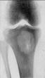 2
2 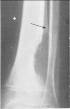 3
3 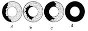 4
4 
Fig.11.9 1 – central destruction; 2 – marginal destruction; 3 – types of sequester (a – cortical, b– central, c – penetrable, d – total); 4 – types of periosteitis (a – linear, b– striped, c– fimbriated, d – lacy, e – needle, f –).
Paraostosis (see fig 11.10)- is stratification of bone tissue, resulting from calcification of adjoining soft tissues (fascia, muscles and enc.).
| BALLON EXPANSION | BLASTOMATOSIS |
| Increase of bone volume with decrision of bone substance in a unit of bone volume. Diameter of bone is wider due to pressure from within of benign growth (cysts and other) | It is disorderly formation of bone tissue on the place of destructed bone; the diameter of bone can be increased. |
| The cortical layer is thinned, clear, is not broken. | The cortical layer often is destroied. |
| The periostitis is absent | Periostal reaction is often as spiculated or Codman’s triangle |
1 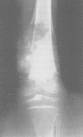 2
2 
Fig.11.10 1 – blastomatosis; 2 – balloon expansion.
The symptoms of joints pathology:
1. The widening of joint space (is the sign accumulation of liquid into a joint cavity).
2. The narrowing of joint space (is the sign of destruction of joint cartilages).
3. The induration of joint capsule (it is becomes visible on sciagram as a result of edema, inflammatory processes, sclerosis and other.
4. The destruction of joint surfaces - characterizes progress of disease, when after destruction of cartilages to destroy of the surface bone. More frequent to destroy surface both bones, the contour of them becomes unclear, irregular, the regional usuration appears sometimes.
5. In the diseases of joints it is possible to arise bone overgrowth (osteophyte) on articular supface of bone.
It is known, that every disease turns to be not a sole symptom. A doctor-roentgenologist distinguishes disease according to all complexes of symptoms – that is by syndrome. This principle of diagnostics facilitates a lot the diagnostic work of a doctor, because there are thousand diseases, and symptoms are considerably less. The selection of syndrome allows to group similar by their displays diseases, that is to divide the great number of diseases in the small number of groups, that allows to make inter-syndrome diagnostics.
For define the leading roentgenologic syndrome one must do three consecutive actions:
1. To make sure that on a sciagram is presented pathological changes (are shown that is to distinguish between norm and pathology.
2. To remember roentgenologic syndromes, met in the diseases of organ explored.
3. To compare a picture exposed on a sciagrams with the standards of all syndromes and attribute the found changes to the definite syndrome.
Syndrome of inflammatory disease of bones:
1. The focus of bone destruction.
2. The bone sequester.
3. The non-malignant periostitis (lamelar, layered, fimbriated, crista-like).
4. Osteoporosis.
5. Osteosclerosis.
The syndrome of inflammatory disease of joints:
1. Widening of joint space
2. Narrowing of joint space
3. Induration of joint capsule
4. Destruction of joints surface of bone
5. Osteonecrosis of epiphysis.
6. Osteoporosis of articular ends of bones (juxtaarticular osteporosis).
Syndrome of bones tumour:
1. Balloon expansion
2. Calcification
3. Blastomatosis
4. Destruction without sequester
5. Malignant periostitis
The signs of syndrome of degenerative-dystrophic disease of joints:
1. the narrowing of joint space
2. the thickening of subchondral bone;
3. the osteosclerosis, especially in the most physically loaded areas;
4. the osteophytes of joint surfaces;
5. the deformation of joint surfaces;
6. the cystic degeneration of joint surfaces.
Syndrome of bones and joints traumatic damage (fractures, luxations): fracture line, displacement of bone fragments, deformation of cortical layer, disorientation of bone structure, deformation of growth plate, dislocation of ossific nucleus, dislocation of articular surface of bones.
Questions for self control:
1. Methods of the bones and joints diseases radio diagnostics.
2. Features of X-ray image|representation| of bones at children|kids|.
Section 12
Date: 2014-12-28; view: 3712