CATEGORIES:
BiologyChemistryConstructionCultureEcologyEconomyElectronicsFinanceGeographyHistoryInformaticsLawMathematicsMechanicsMedicineOtherPedagogyPhilosophyPhysicsPolicyPsychologySociologySportTourism
Cells Vary in Shape, Size and Arrangement
Our own body contains over 100 different kinds of cells. Some of these cells are round. Others, such as the nerve cells, are shaped like long, tangled strings. Cells tend to be spherical. They take other shapes because of cell walls found in most plant cells and in many one-celled organisms or because of attachments to and pressure from other, neighboring cells or surfaces.
Cells vary greatly in size too (Fig. 1.1). Most of the cells that make up a plant or animal body are within a size range of between 10 and 30 micrometers in diameter. The yolk of a single ostrich egg is one of the largest cells known. It has a circumference of nearly 8 centimeters. In contrast, the bacteria that naturally live deep inside your body are microscopic. And the most metabolically active cells are usually small too. The ostrich egg cell is 800,000 times bigger than the bacterial cells.
The living arrangements of cells also vary.
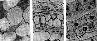
Fig. 1.1. The cells of a plant (left), a rat (center),
and a human being (right) appear to be quite different
Organization of Cells
Every cell is a self-contained and at least partially self-sufficient unit surrounded by an outer membrane that controls the passage of materials in and out of the cell and so makes it possible for the cell to differ biochemically and structurally from its surroundings. All cells also, at least at some time in their existence, contain control centers or nuclei. Many have a variety of internal structures, the organelles (Fig. 1.2) which are similar or identical from one cell to another throughout a wide range of cell types. All are composed of the same remarkably few kinds of atoms and molecules.
The cell membrane covers the entire surface of the cell. New materials for growth and energy enter the cell through tiny holes in this membrane. Waste products leaving the cell must also go through the cell membrane. Others must be pumped in and out. The pumping process uses some of the cell’s energy.
The cytoplasm is a jellylike substance which fills most of the cell. Many of building materials of a cell are manufactured and stored here.
A large, round nucleus is found somewhere in the cytoplasm. As the «control center» of the cell, the nucleus contains coded instructions for all of the activities of a cell. These coded instructions are stored on special structures called chromosomes. Chromosomes are seen when a cell is reproducing.
| Vacuolar membrane Large central vacuole Tiny channel connecting cytoplasm of neighbouring cells Smooth endoplasmic reticulum (lacks ribosomes) | 
| Cell wall Cell membrane Mitochondrion Chloroplast Nuclear membrane Nucleus Pore in membrane Nucleolus Rough endoplasmic reticulum Ribosome Golgi apparatus Secretory vesicle |
| Fig. 1.2. Ultrastructure of generalized plant cell |
A nuclear membrane covers the nucleus of the cell. It regulates the passage of materials in and out of the nucleus.
Sausage-shaped bodies in the cytoplasm are mitochondria. Single cells may contain hundreds of these structures. Mitochondria are two-membraned organelles including all eucaryotic cells. They are round-shaped, stick-shaped, threads-shaped bodies (0.5-10 nm long). Sometimes they branch out. Their number is diverse: 1-100000 and more, depending on metabolic activity. Superficial apparatus of mitochondrion consists of two membranes: outer and inner ones. The outer one is smooth; it separates the mitochondrion from the hyaloplasma. The inner one formats bends inside the mitochondrion, which look like a tube holes. They can be distributed about the longitudinal axis of the mitochondrion in different ways; they are branched out very often. The outer and inner membranes are separated by the cavity 10-20 nm wide. ATP-soma, mushroom-like holes, containing enzyme complex, needed for ATP’s synthesis are on the surface of the inner membrane. The inner space is filled with half-liquid substance-matrix. The molecules of DNA and RNA are in it. The proteins, constituting the content of the inner membrane, are synthesized there.
The principal mitochondrion’s function is the synthesis of ATP, occurring owing to the energy educed during oxidizing the organic compounds. Mitochondria are commonly called the “power houses of the cell”. They trap the energy that results when food is broken down.
A network of canals or a system of cavities, which looks like microscopic canals and thickenings, is called the endoplasmic reticulum. These canals lead from the nuclear membrane to the cell membrane. The diameter of the canals is more than 50 nm, their thickenings are called cisterns. Life scientists think that the endoplasmic reticulum may be involved in the transport and storage of substances. Two kinds of endoplasmic reticulum are known: rough and smooth ones. The proteins, coming into the cavities of the rough endoplasmic reticulum, can be accumulated there, mature and get secondary, tertiary and quaternary structures, attach non- proteins particles etc. Moreover, the rough endoplasmic reticulum takes part in the synthesis of the cell’s membranes. The smooth endoplasmic reticulum has no ribosomes on its membranes.
Ribosomes are the tiny and dense dots located on the membranes of the rough endoplasmic reticulum, which form the complexes with mRNA-polyribosomes during the biosynthesis of protein. They also distribute the synthesized proteins between different parts of the cell. The actual building blocks of the cell are made on the surfaces of these very tiny structures.
The Golgi body is a heap of the smooth cisterns covered with membranes; within them we can see bubbles and canals. The cisterns are polar as a rule: the bubbles come to one pole, merge with them, giving away their contents, then, filled with different substances, separate from the other pole of the cistern and transport these compounds into other parts of the cell or push them out. Specifically Golgi body takes part in the creation of lysosome containing hydrolytic enzymes synthesized on the membranes of the rough endoplasmic reticulum. All eucaryotic cells contain Golgi body but its structure can vary in different organisms. The functions of the Golgi body are diverse. Some substances are accumulated and changed there, covered with membranes and secret later. The substances, getting cisterns, are sorted according to the chemical composition and function there. In the next cisterns, sorted molecules mature, separate from the Golgi body. The bubbles, having separated, can merge with the membranes of the endoplasmic reticulum or with plasmatic membrane pushing their components out. Some of them come to other organelles and give their contents. Golgi body takes part in the building of the plasmatic membrane and other cell membranes. During division of the cell, Golgi body decomposes into separate structural units distributed among daughter cells accidentally.
Lysosomes are the bubbles, 100- 800 nm in diameter, surrounded by the membrane. They contain different hydrolytic enzymes, available to the dissolution of the organic compounds, and ensure the processes of intracellular nutrition. Lysosomes are formed in the Golgi body. The enzymes, constituting them, are synthesized on the membranes of the rough endoplasmic reticulum, then they are transported to the cisterns of Golgi body and separate as bubbles surrounded by the membrane. There are lysosomes of different types in the cell. Primary lysosomes are formed due to Golgi body merging with pinocytic or phagocytic bubbles; they form the secondary lysosomes. The enzymes of the primary lysosomes are activated after merging and their contents is digested. If in the secondary lysosomes the compounds or the microorganisms are decomposed not entirely, they are transformed into residual bodies. Some of them are pushed out, others stay in. One more type of lysosomes-autolysosome - takes part in the digesting, separates components of the cell, whole cells or their groups.
Vacuoles are the cavities in the cytoplasm surrounded by the membrane and filled with the liquid. There are different types of vacuoles in the cells of eucaryote. Vacuoles of the plant cells are formed from the bubbles separated from the endoplasmic reticulum. The small vacuoles merge into large ones, taking almost entire volume of the plant cell. They are filled with the cell’s sap-aqueous solution of organic and inorganic compounds, including the products of exchange or pigments. The functions are diverse: they support the turgor pressure favouring to the preserving of the contain cell’s shape; soluble reserve nutritious substances and the toxic exchange products are accumulated there. Contractive vacuole of the fresh-watered one-celled animals and water-plants are formed from the elements of the Golgi body. They regulate osmotic pressure, take part in taking out the dissolved metabolic products, favour entering oxygen and water into a cell.
Plastids are two-membraned organelles of the plants’ cells and of some animals. They are diverse according to the shape, size, color and the structural peculiarities. There are 3 kinds of them: chromoplasts, leucoplasts and chloroplasts.
Chloroplasts are the plastids colored green due to chlorophyll pigment. As a rule, they are long-shaped (5-10 nm), their number is diverse in the cells. There is a space between the outer and inner membranes of chloroplasts 20-30 nm wide. The inner membrane formats folder-bends into the matrix: lamellas and telacoids. The former looks like long folds while the letter-like sacks gathered into a heap of 50 or more. It is called grana, containing the main photosynthetic pigment–hlorophyll, and also auxiliary – arotinoids. The main function of chloroplasts is photosynthesis.
Chloroplasts have a definite state of autonomy in the cell. They have own hereditary information, protein-synthesized body, connecting ribosomes and all kinds of RNA, synthesizing specific proteins constituting their membranes. They reproduce by division.
Leucoplasts are colorless plastids of different shape, have no developed lamella system. There are DNA, ribosomes, enzymes, providing synthesis and hydrolysis.
Chromoplasts are the plastids of different colors-yellow, red etc. They color leaves and fruits; color depends on different pigments. The inner membrane’s system is absent.
Date: 2014-12-22; view: 1934
| <== previous page | | | next page ==> |
| BASICS OF ORGANISATION OF LIFE | | | Plant, Animal and Bacterial Cells |