CATEGORIES:
BiologyChemistryConstructionCultureEcologyEconomyElectronicsFinanceGeographyHistoryInformaticsLawMathematicsMechanicsMedicineOtherPedagogyPhilosophyPhysicsPolicyPsychologySociologySportTourism
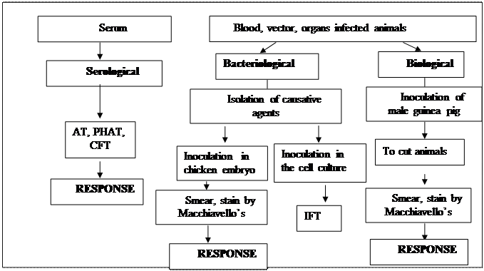
Microbiological research of the Ricketsia disease
LABORATORY DIAGNOSTIC OF MYCOPLASMA AND CHLAMIDIA DISEASES
Actuality.There are over 150 species in the class cell wall-free bacteria known as Mollicutes. In humans, four species are of primary importance: Mycoplasma pneumonia, Mycoplasma hominis, Mycoplasma genitalium and Ureaplasma urealiticum. It causes pneumonia, urethritis, postpartum fever, and other infections.
Primary objectives: to be able to conduct and evaluate the microbiological diagnostics of Mycoplasma and Chlamidia infections.
.
QUESTIONS FOR DISCASSION
1. General description of bacteria of the family Mycoplasma and Chlamidia.
2. Microbiological diagnosis of the diseases caused by Mycoplasma and Chlamidia.
3. Epidemiology and pathogenesis of the diseases caused by Mycoplasma and Chlamidia. Features of immunity in such diseases.
4. Principles of prophylaxis and treatment of the diseases caused by Mycoplasmas and Chlamidia.
PROCEDURE OF PRACTICAL SESSION
Task 1. To inoculate the sputum from patients with candidiasis pneumonia on the media.
Task 2. Examine microscopically the smears from the patient's materials fill in the protocol.
Task 3. Study the growth of the Mycoplasma hominis on the solid media.
Task 4. Study and write to the protocol information about immunobiological preparates for diagnostic of the Chlamidia infection.
RECOMMENDATIONS FOR PRACTICAL WORK
Task 1. You must inoculate 0.1 ml of the diluted (1:100) sputum on the Petri dish with potatoes media. The material is spreaded on the surface of the media for to do growth that like lawn. After that, Petri dish will putted for incubation.
Task 2.
 IFT.
IFT.
The arrow shows the inclusion of the Chlamidia in cytoplasm of the epithelial cells.
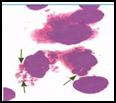
Chlamydiae have distinctive staining properties (similar to those of rickettsiae). Elementary bodies stain purple with Giemsa stain in contrast to the blue of host cell cytoplasm. The larger, noninfective reticulate bodies stain blue with Giemsa stain.
Task 3.

Mycoplasma hominis has been sealing to prevent evaporation, isolated colonies measuring 20-500 μm can be detecting with a hand lens. These colonies are round, with a granular surface and a dark center typically buried in the agar. They can be subculturing by cutting out a small square of agar containing one or more colonies and streaking this material on a fresh plate or dropping it into liquid medium.
Task 4.
Dry chlamidia diagnosticum for CFT is used for diagnostic in complement fixation test for detection specific antibody. It contains killed chlamidiа.
Addition 1
Date: 2016-01-14; view: 3554
| <== previous page | | | next page ==> |
| Zdrodovsky staining. | | | Laboratory diagnostics of Mycoplasma and Chlamidia diseases |