CATEGORIES:
BiologyChemistryConstructionCultureEcologyEconomyElectronicsFinanceGeographyHistoryInformaticsLawMathematicsMechanicsMedicineOtherPedagogyPhilosophyPhysicsPolicyPsychologySociologySportTourism
LABORATORY DIAGNOSTICS OF DIPHTERIA AND WHOOPING COUGH (PERTUSSIS)
Actuality: Corynebacterium diphtheriae is the most important member of the group, it can produce an exotoxin that causes diphtheria in humans. Bordetella pertussis, a highly communicable and important pathogen of humans, causes whooping cough (pertussis).
Primary objectives: to be able to conduct and evaluate the microbiological diagnosis of the diphtheria and pertussis (whooping-cough).
QESTIONS FOR DISCASSION
1. Biological properties of diphteria and pertussis causative agent.
2. Pathogenesis of diphtheria and pertussis in humans.
3. Features of antidiphtherial and antipertussis immunity.
4. Laboratory diagnostics of diphtheriae and pertussis.
5. Specific prophylaxis and treatment of the disease.
PROCEDURE OF PRACTICAL SESSION
Task 1. Prepare the smears from pure culture C. diphtheriae Gram stain, Neisser stain and Löffler stain.
Task 2. Study the growth of Corynebacteriun diphteriae cultures on Löffler nutrient medium and Buchin's medium.
Task 3. Study the diphtheria bacillus cultures toxigenicity in gel-precipitation assay.
Task 4. Study the differentiative properties of corynebacteria.
Task 5. Study the test of cistinase (Pizu test).
Task 6. Study the test of urease (Zaks test).
Task 7. Read the indirect hemagglutination test, make a conclusion about antidiphtheritic immunity.
Task 8. Prepare the smears from pure culture of Bordetella pertussis, stain by Gram method.
Task 9. Study the differentiative features of bordetella.
Task 10. Read the agglutination test with patient's pair sera and whooping cough diagnosticum. Make the conclusion
Task 11. Describe the immunobiological preparates for treatment and prophylaxis of diphtheriae and pertussis.
RECOMMENDATIONS FOR PRACTICAL WORK
Task 1.
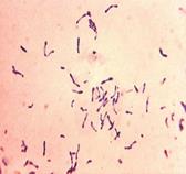 Diphtheria bacteria are Gram-positive, pleomorphic, often club-shaped rods. The individual cells tend to group in V, Y, or palisade arrangements.
At Löffler stain Corynebacterium spp. is blue. In both cases of valutins grain, painted more intensively than the central part of the cells (phenomenon of metachromasia). Neisser staining reveals the polar bodies (polyphosphates stored at one end of the rod).
 Task 2.
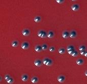
The usual media employed for cultivation of the diphtheria bacillus are Löffler‘s serum slope and tellurite blood agar. Diphtheria bacilli grow on Löffler's serum slope very rapidly and colonies can be seen in 6-8 hours, long before other bacteria grow. Colonies are at first small, circular white opaque discs but enlarge on continued incubation and may acquire a distinct yellow tint.
Tellurite (0.4 %) inhibits the growth of most other bacteria, acting as a selective agent. Diphtheria bacilli reduce tellurite to metallic tellurium, on the tellurite medium and colonies became grey or black color. The growth of diphtheria bacilli may be delayed. It may take 2 days to appear.
Buchin's medium consists of agar, sprat hydrolysat, sodium chloride, chincsolum, glucose, water-blue indicators. It is prepared from powder, according to the label instructions. It is boiled for 2-3 minutes and cooled to 50°C, after that 50 ml of defibrinated blood (rabbit or human) is added. The prepared medium is dark blue. On this medium C. diphtheriae forms dark blue colonies, diphtheroids - light blue ones.
Task 3.
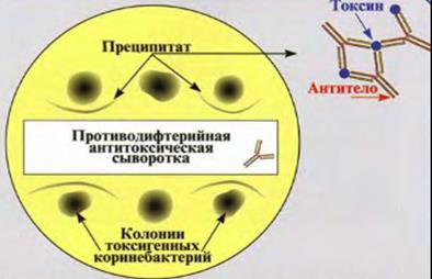
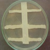 The gel precipitation test is based on the interaction of homologous antibodies and antigens in gel and the formation of visible bands of precipitation. Because of counter-diffusion into gel, the antibodies and antigen form immune complexes (aggregates) visualized in the form of opalescent white bands.
When several antigens diffusing irrespective of each other are present, the number of bands corresponds to the number of antigens. Serologically homogeneous antigens form precipitation bands which merge with each other, whereas bands of heterogeneous antigens cross each other. This property permits determination of the homogenecity of the antigenic structure of various objects tested.
Components of the gel precipitation test are gel, antigen, and antibodies. For quality control of the gel precipitation test, the test system comprised of known homologous antibodies and antigens is utilized. The antigen used in a precipitation test should be concentrated, while the sera (from patients or immunized animals) should be of a high titer.
Procedure. In a solidified agar cut the wells and remove the agar from them with a pasteur pipette. Into 1 series of walls place serum, into the other - antigens and put the slides into a humid chamber for several days. In reading the results of the reaction, compare the localization and nature of precipitation lines in the test and control wells. To measure the levels of the antigen and antibodies, study their multiple dilutions. The precipitation test in gel is widely used in the diagnosis of diseases caused by viruses, rickettsiae, and bacteria producing exotoxins. It has become of great practical significance with regard to determining the toxigenicity of Corynebacteria diphtheria.
|
|
|
|
|
Date: 2016-01-14; view: 1850
| <== previous page | | | next page ==> |
| Laboratory Diagnostics of the Anaerobic Infections | | | Task 4. The differentiating properties of corynebacteria |