CATEGORIES:
BiologyChemistryConstructionCultureEcologyEconomyElectronicsFinanceGeographyHistoryInformaticsLawMathematicsMechanicsMedicineOtherPedagogyPhilosophyPhysicsPolicyPsychologySociologySportTourism
Age-related features of fractures
Children`s fractures:
- subperiosteum fracture - on the type of “green twig” (integrity of cortical layer is disintegrated on the one side, and from the opposite side the disentegration are not present), a periosteum is left intact, displacements are not present.
- Impacted fractures - when the fracture line appears how area decreased radiolucent – radiopaque line fracture.
- epiphysiolysis, osteoepyphysiolisis - the line of fracture passes trought area of epiphyseal cartilage. Radiological signs: displacement of epiphyseal centers of ossification in relation to metaphysis. Often epyphysiolisis is combined with the metaphysis fractures - osteoepyphysiolisis.
Specific feature of elderly age fractures.
As a result osteoporosis the fractures can arise at an action of insignificant injuring factors. Thus there often are the multifragmental, and sometimes multiple fractures, delayed union (hypoporosis).
Healing of fracture (see fig 14.6) result from formation of a callus.
Ø The connective tissue callosity - appears in the site of the fracture during 7-10 days after a trauma.
Ø The osteoid callosity is the transition of connecting tissue in osteoid one, with formation of osteoid trabecoules.
Ø A bone callosity is the process of calcification of osteoid tissue, takes from 3-4 weeks to 8 months (depending on the size of bone, features of the fracture and the patient age). In 4-8 months the fracture line disappears, fragments consolidation comes.
Ø Involution of callosity - a callosity resorbs partly, beginning from periphery, the bone structure renewels during 1-2 years.
Fig.14.6 Bone callosity
Complication of fractures: vicious union (malunion), excessive callus, false joint, bone defect, traumatic osteolisis, simple necrosis, traumatic osteomielitis and traumatic myositis ( see fig 14.7).
1 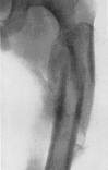 2
2  3
3 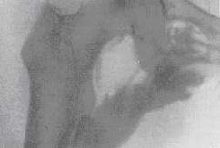
Fig.14.7 1 – malunion, excessive callus; 2 –false joint; 3 – myositis ossificans.
Dislocations (see fig 14.8)
Dislocation is violation of congruention of a joints surfaces (articular surface).
- complete dislocation - if joint surfaces do not touch;
- incomplete dislocation - when the joint surfaces of bones touch partly.
1 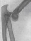 2
2 
Fig.14.8 1 – complete traumatic dislocation of elbow joint (sciagram in lateral view); 2 – fracture-dislocation carpal articulation (wrist joint) (two-dimension sciagram)
Dislocations include the following types: congenital and acquired. Among acquired ones selects: traumatic, pathological - in destruction of elements of the joint, distensional - at accumulation of liquid in the bag of a joint, ordinary dislocation.
Date: 2014-12-28; view: 2202
| <== previous page | | | next page ==> |
| The radio diagnostics of the musculosceletal system’s tumours | | | Английский язык Для студентов-юристов |