CATEGORIES:
BiologyChemistryConstructionCultureEcologyEconomyElectronicsFinanceGeographyHistoryInformaticsLawMathematicsMechanicsMedicineOtherPedagogyPhilosophyPhysicsPolicyPsychologySociologySportTourism
Pleurisy
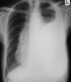
Pleurisy (see fig 16.6) is a disease of pleura, which arises up more frequently as the second process is complication of pneumonia, tuberculosis, other diseasees of lungs, heart, blood and others like that. Distinguish a dry and exudative pleurisy.
Development of dry (fibrinous) pleurisy is a conditioned productive inflammation of pleura. Patients mark a cough, increase of body temperature, pain in breasts, which increases during cough. Roentgenological limited a dry pleurisy is not shown up. Widespread dry pleurisy is accompanied by increasing of interlobar and costal pleura (to 1 sm), by the decline of radiolucency lungs, unclearness of contours of costal-diaphragm recesses. Sometimes in a pleura cavity by ultrasonic examination it is succeeded to find out the two-bit of liquid.
Presence of liquid in a pleura cavity is better to find out in lateropositsion of the patient on to t sides. Sciagraphy finds out in a pleura cavity a liquid by volume of more than 0,5l ultrasound - more than 20 ml.


Fig.16.6 Exudative pleurisy on a sciagram and ultrasound
1 a liver; 2 a diaphragm; 3 exudate; 4 a visceral pleura
Ran across exudative pleurisy characterise more expressed clinical signs. On sciagrams in the anterior view of accumulation of exudate in a pleura cavity at vertical position of patient notedly a darkening of lateral costal-diaphragmal recess, or three-cornered shade in the lateral department of the inferior pulmonaris field. If a high bound of the shade is at the level of body of the 5th rib, amount of liquid is approximately 1l, at the level of the 4th rib-1,5l, 3th rib –2l. The greater amount of exudate anymore displaces the organs of mediastinum to the healthy side.
Pleura accretions can divide a pleura cavity into the separate isolated departments, forming encapsulation exudative pleurisies.
Shades of such pleurisies are not displaced at the change of the body position, their contours are clear and protuberant, they resolve slowly. The place of location is costal, costal-diaphragmal, costal-vertebral, apex, diaphragmal, mediastinal, interlobar encapsulation pleurisies.
Radiodiagnosis of the tuberculosis
Date: 2014-12-28; view: 1554
| <== previous page | | | next page ==> |
| Functional symptomes | | | Tuberculosis |