CATEGORIES:
BiologyChemistryConstructionCultureEcologyEconomyElectronicsFinanceGeographyHistoryInformaticsLawMathematicsMechanicsMedicineOtherPedagogyPhilosophyPhysicsPolicyPsychologySociologySportTourism
Figure 1. Sound-cue finger tapping task.
Subjects
Twenty-six children with a diagnosis of migraine who had acute migraine attack (20 girls, 6 boys; mean age 14.7±1.9 years) were recruited from our Headache Clinic (see table 1). The participants were pre-screened by pediatric neurologists specialized in headache at our Headache Clinic at CCHMC. If a participant met the criteria and was interested in our MEG study, a researcher would explain the research protocol and obtain written informed assent and consent forms from the participant and her/his parents. The research protocol, assent and consent forms were formally approved by the Institutional Review Board (IRB) at CCHMC. Inclusion criteria for children with migraine was: clinically diagnosed migraine and met diagnostic criteria defined in the International Classification of Headache Disorders, 2nd Edition [12]. Healthy controls were recruited to match the patients for age and gender and met inclusion criteria of: (1) healthy without history of neurological disorder, migraine or brain injury; (2) age-appropriate hearing, vision, and hand movement. Exclusion criteria for all subjects were: (1) inability to remain still; (2) presence of an implant such as a cochlear implant device; a pacemaker; or a neuro-stimulator containing electrical circuitry, generating magnetic signals, or having other metal that could produce visible magnetic noise in the MEG data; (3) noticeable anxiety and/or inability to readily communicate with personnel operating the MEG system. The MEG studies were performed prior to initiation of treatment.
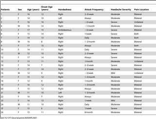
Table 1. Demographic and Clinical Features of Children with Migraine.
doi:10.1371/journal.pone.0050095.t001
- Download:PowerPoint slide | larger image (108KB PNG) | original image (484KB TIFF)
Motor Task
All subjects performed a brisk left or right index finger tapping immediately after hearing a sound cue (500 Hz, 30 milliseconds (ms) square tone). Subjects were instructed to press a response button with the index finger that was ipsilateral to the tone (see Figure 1). The eyes were open and fixed to an arbitrary target during the paradigm. A trigger was sent to the MEG system from the response box when the button was pressed. The stimuli consisted of 200 trials of square tones, 100 trials per ear, and were presented randomly through a plastic tube and earphones. The time window for finger movement was 3000 ms; the inter-stimulus interval was 0–1000 ms, which varied from 0 to 1000 ms randomly. Therefore the time between two consecutive auditory cues was 3000–4000 ms. The stimulation presentation and response recording were accomplished with the BrainX software [13], which was a software package based on DirectX (Microsoft Corporation, Redmond, WA, USA).

Figure 1. Sound-cue finger tapping task.
A tone is sent to the participant’s left or right ear in a randomized order: The participant is instructed to press a button on her/his left side when the tone is sent to the left ear; the participant is instructed to press a button on her/his right side when the tone is sent to the right ear. Each button will send a unique signal to the MEG system in real-time and the MEG system will record and store the unique signals to the MEG dataset for analysis of movement-related neuromagnetic responses.
doi:10.1371/journal.pone.0050095.g001
- Download:PowerPoint slide | larger image (1.38MB PNG) | original image (1.5MB TIFF)
MEG Recordings
The neuromagnetic signals were recorded in a magnetically shielded room (Vacuum-Schmelze, Hanau, Germany) using a whole head CTF 275-Channel MEG system (VSM MedTech Systems Inc., Coquitlam, BC, Canada) in Cincinnati Children’s MEG Center prior to clinical treatment for the participants. This magnetic shielded room was designed to reduce environmental magnetic noise. Before data acquisition commenced, three electromagnetic coils were attached to the nasion, left and right pre-auricular points of each subject. These three coils were subsequently activated at different frequencies for measuring each subject’s head position relative to the MEG sensors. Each subject was comfortably positioned in the supine position with arms resting on either side, during the entire procedure. The sampling rate of the MEG recording was 6000 Hz per channel. An acquisition window was set to 3000 ms per trial, with 2000 ms pre-trigger and 1000 ms post-trigger. The data were recorded with a noise cancellation of third order gradients and without on-line filtering. Subjects were asked to remain still. If head movement during a recording was beyond 5 mm, that dataset was indicated as “bad” and an additional trial was recorded.
Date: 2016-03-03; view: 964
| <== previous page | | | next page ==> |
| Information on what experts propose as alternative solutions | | | Figure 6. Magnetic source activation of high-gamma oscillations elicited by right finger movement. |