CATEGORIES:
BiologyChemistryConstructionCultureEcologyEconomyElectronicsFinanceGeographyHistoryInformaticsLawMathematicsMechanicsMedicineOtherPedagogyPhilosophyPhysicsPolicyPsychologySociologySportTourism
RECOMMENDATIONS FOR PRACTICAL WORK
|

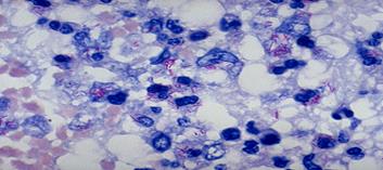
Tubercle bacilli are more strongly acid-fast the other members of the acid-fast group and have a characteristic beaded applesauce. Both Gram's stain and acid-fast stain depend on the integrity of the cell wall. Broken or disintegrated bacilli or their parts are neither gram-positive nor acid-fast.
Ziehl-Neelsen Stain Procedure
1. Fix the smear.
2. Flood the slide with carbolfuchsin, steam gently for 5 minutes over low flame, and do not allow drying and adding more stain if necessary. Cool. Alternatively, carbolfuchsin-containing phenol and alcohol (cool) may be used without heat.
3. Apply 90 % alcohol containing 3 % to 5 % HC1 until all but the thickest parts of the smear cease to give off color (approximately 1 to 3 minutes). Wash.
4. Stain 1 minute with methylene blue. Wash.
5. Examine smear using the oil-immersion lens of the light microscope.
Task 2.
|
Task 3.
|

A simple medium containing only eggs, malachite green and coconut water has been reported to be a useful and cheap alternative to the Lowenstein-Jensen medium.
Among the several liquid media described, Dubos', Middlebrook's, Proskauer and Beck's, Sula's and Sauton's media are the more common. On solid media M. tuberculosis forms dry, rough, raised, irregular colonies with a wrinkled surface. They are creamy white initially, becoming yellowish or buff colored later. They are tenacious and not easily emulsified. The colonies of M. bovis are in comparison, flat, smooth, moist and white, breaking up easily when touched. Liquid media are not generally employed for routine cultivation but are used for sensitivity tests chemical tests and preparation of antigens and vaccines. In liquid media without dispersing agents, the growth begins at the bottom, creeps up the sides and forms a prominent surface pellicle that may extend along the sides above the medium. Diffuse growth is obtained in Dubos' medium containing a detergent Tween-80 (sorbitan monooleate). Virulent strains tend to form long serpentine cords in liquid media, while avirulent strains grow in a more dispersed fashion. The cord factor by itself is not responsible for virulence. It is present in some nonpathogenic species of mycobacteria as well. Colonial morphology may be modified by the presence of bacteriophage in the strain. Tubercule bacilli may also be grown in chick embryos and in tissue culture.
Task 4.
For identification of the selected cultures of tuberculosis causative agents and differentiation them from other types of Mycobacteria utillize a lot of signs. Basic from them resulted in a table.
The differential properties of Mycobacteria for identification
| Time of grow, days | Catalase, 68°C | Urease | Nicotin-amidase | Niaci-nase | Nitratere-ductase | Pigmet | |
| M. tuberculosis | 12 - 25 | - | ± | + | + | + | - |
| M. bovis | 24 - 40 | - | + | - | - | - | - |
| M. africanum | 31 - 42 | - | + | + | - | - | - |
| M. kansasii | 10 -20 | + | + | + | - | + | + |
| M. avium | 10 - 12 | + | - | + | - | + | - |
| M. smegmatis | 3 - 5 | ± | + | + | - | + | - |
Most essential sign of M. tuberculosis is a niacine test and ability to synthesize plenty of nicotin acid (Niacinum). Catalase activity is relatively weak and losts at 68°С.
Task 5.
The determination of MBT sensitivity to antimycobacterial preparations is of paramount importance for the treatment tactics, correction of antimycobacterial therapy and the illness prognosis. MBT sensitivity to antituberculous preparations is defined by the preparation minimum concentration, which inhibits MBT growth on the nutritive medium. MBT are considered to be sensitive to either preparation if less than 20 colonies have grown in a test-tube, with abundant rowth in the control. The culture is supposed to be stable if more than 20 colonies have grown. MBT are considered to be stable if they grow at the concentrations of the preparation in 1 ml of the nutrient medium: for isoniazidum – l mkg, rifampicinum - 20 mkg, streptomycini - 5 mkg, ethambutolum - 2 mkg, all other preparations - 30 mkg.
Medicinal resistance of tuberculosis causative agents is determined by the method of the serial delutions before the beginning of treatment, in 3 months and farther at continuation of selection of tubercular sticks through each 6 months. It is done by growing of cultures on medium with the different concentration of tuberculostatics. Most widespread followings methods of determination of medicinal stability of mycobacteria: cultivation on the dense media of Löwenstein-Jensen; microcultivation on glasses by Price method; deep sowing in semisynthetic agar media. In test tubes which contain the different concentration of preparations, and in one control (without tuberculostatics) suspension of pure culture (500 ml of microbal bodies is in 1 ml) is inoculated. A culture is considered sensible, if in a test tube > 20 colonies grew is massive growth in control. If more than 20 colonies grew, a culture is considered steady. Resistance of this culture is expressed the that maximal concentration of antibacterial preparation, at which yet there is growth, close growth in a control test tube (table.)
Scale of estimation of resistance of mycobacteria to medicinal preparations
| Preparate | Resistent at growth on media, which contain preparation, mcg/ml | |
| dense | liquid | |
| Streptomycini sulfas | ||
| Izoniazidum | ||
| Rifampicinum | ||
| Ethambutolum | ||
| Canamycini sulfas | ||
| Ethionamide | ||
| Biomycin |
Task 6.
Isoniazidum is the principal representative of hydrazide isonicotine acid group (HINA), the most effective among all antimycobacterial drugs, strictly specific only against MBT, penetrates through cell and tissue membranes and through haematoencephalic barrier well
Rifampicinum is a semisynthetic antibiotic with a wide action spectrum. The drug possesses an expressed bacteriostatic activity to MET, which are distributed extracellularly and in the cells (intracellularly), as well as to the ones that multiply quickly and slowly.
Task 7.
Tuberculinis an allergen that used for tuberсulin skin test or Mantoux. It is utilized for determination of infectation population, mass inspection, tuberculosis of children and teenagers, selection of persons, which need to conduct revaccination, check up its efficiency. In addition, this test is utilized with the purpose of diagnostics of tuberculosis and determination of activity of infectious process. For raising tests utilize a tuberculin. It is prepared from mixture of cultures of human and bovine types Mycobacteria, growing in glycerin clear both. A culture is sterilized current steam 30 minutes, evaporate, filter through a bacterial filter and pour out in ampoules. Preparation is introduced subcuteniously.
Lepromin (allergen) is prepared from sterilized in the autoclave of the staggered fabrics that contain plenty of mycobacteria. Lepromin introduced intracuteniously in middle third of forearm in a volume 0,1 ml. In 48 hours in positive cases to form spot of eritema or papula (reaction of Fernandesa) of considerably, later (1-2 months) can appear tubercle, often with necrosis (reaction of Mitsuda's). In patients with lepromatous form and healthy people an allergic reaction is negative, in patients with tuberculoid form the test is positive. The test does not have a diagnostic value, it is utillized only for determination of clinical form of disease and prognosis.
Erythrocytes tuberculosis diagnosticum is utilized in serum diagnostics of tuberculosis for raising of PHAT, which is put with the purpose of exposure of specific antibodies in the serum of blood of patient. On-the-spot sheepskin red corpuscles the phosphatides antigen of M. tuberculosis is adsorbed. Serum method behaves to the additional methods of diagnostics of tuberculosis. A positive reaction is marked at an active tubercular process, and at infected mycobacterias of tuberculosis and after a vaccination.
Task 8.
BCG (bacillus Calmette-Guerin) is living or attenuated, liophilic dried up culture unpathogenic strain of M. tuberculosis, was found by the French scientists Calmette and Guerin. It is used for the active specific prophylaxis of tuberculosis. It is plugged in the calendar of inoculations. It is contra-indicated for people with violation of cellular link of immunity.
BCG-М difference from previous preparation is only in an amount mycobacterias (less than in 2 times). It intended for an active specific prophylaxis for hyposthenic children.
Addition 1
Date: 2016-01-14; view: 3490
| <== previous page | | | next page ==> |
| Laboratory Diagnostics of the Diphtheriae and Pertussis | | | Laboratory Diagnostics of the tuberculosis and leprosy |