CATEGORIES:
BiologyChemistryConstructionCultureEcologyEconomyElectronicsFinanceGeographyHistoryInformaticsLawMathematicsMechanicsMedicineOtherPedagogyPhilosophyPhysicsPolicyPsychologySociologySportTourism
Ultrasound Imaging
Ultrasound imaging, also called ultrasound scanning or sonography, involves exposing part of the body to high-frequency sound waves to produce pictures of the inside of the body. Ultrasound examinations do not use ionizing radiation (as used in x-rays). Because ultrasound images are captured in real-time, they can show the structure and movement of the body's internal organs, as well as blood flowing through blood vessels.
Ultrasound imaging is a noninvasive medical test that helps physicians diagnose and treat medical conditions.
Ultrasound imaging of the breast produces a picture of the internal structures of the breast.
Doppler ultrasound is a special ultrasound technique that evaluates blood flow through a blood vessel, including the body's major arteries and veins in the abdomen, arms, legs and neck.
During a breast ultrasound examination the sonographer or physician performing the test may use Doppler techniques to evaluate blood flow or lack of flow in any breast mass. In some cases this may provide additional information as to the cause of the mass.
Some common uses of the procedure
Determining the Nature of a Breast Abnormality
The primary use of breast ultrasound today is to help diagnose breast abnormalities detected by a physician during a physical exam (such as a lump or bloody or spontaneous clear nipple discharge) and to characterize potential abnormalities seen on mammography or breast magnetic resonance imaging (MRI).
Ultrasound imaging can help to determine if an abnormality is solid (which may be a non-cancerous lump of tissue or a cancerous tumor) or fluid-filled (such as a benign cyst) or both cystic and solid. Ultrasound can also help show additional features of the abnormal area.
Doppler ultrasound is used to assess blood supply in breast lesions.
Supplemental Breast Cancer Screening
Mammography is the only screening tool for breast cancer that is known to reduce deaths due to breast cancer through early detection. Even so, mammograms do not detect all breast cancers. Some breast lesions and abnormalities are not visible or are difficult to interpret on mammograms. In breasts that are dense, meaning there is a lot of ducts, glands, fibrous tissue and less fat, many cancers can be hard to see on mammography.
Many studies have shown that ultrasound and magnetic resonance imaging (MRI) can help supplement mammography by detecting small breast cancers that may not be visible with mammography. MRI is more sensitive than ultrasound in depicting breast cancer, but MRI may not be available to all women. If screening MRI is performed, then screening ultrasound is not needed, though ultrasound may be used to characterize and biopsy abnormalities seen on MRI. When ultrasound is used for screening, many more abnormalities that may require biopsy are seen than are seen with mammography or MRI. These abnormalities usually are not cancer (false positives), and this limits its usefulness.
Ultrasound can be offered as a screening tool for women who:
-are at high risk for breast cancer and unable to undergo an MRI examination.
-are pregnant or should not be exposed to x-rays (which is necessary for a mammogram).
Ultrasound-guided Breast Biopsy
When an ultrasound examination reveals a suspicious breast abnormality, a physician may choose to perform an ultrasound-guided biopsy. Because ultrasound provides real-time images, it is often used to guide biopsy procedures. An ultrasound exam will usually need to be performed before the biopsy in order to plan the procedure and to determine if this method of biopsy can be used.
What does the equipment look like?
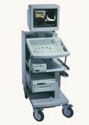
Ultrasound scanners consist of a console containing a computer and electronics, a video display screen and a transducer that is used to do the scanning. The transducer is a small hand-held device that resembles a microphone, attached to the scanner by a cord. The transducer sends out inaudible high frequency sound waves into the body and then listens for the returning echoes from the tissues in the body. The principles are similar to sonar used by boats and submarines.
The ultrasound image is immediately visible on a video display screen that looks like a computer or television monitor. The image is created based on the amplitude (strength), frequency and time it takes for the sound signal to return from the area of the patient being examined to the transducer and the type of body structure the sound travels through.
How does the procedure work?
Ultrasound imaging is based on the same principles involved in the sonar used by bats, ships, fishermen and the weather service. When a sound wave strikes an object, it bounces back, or echoes. By measuring these echo waves, it is possible to determine how far away the object is and its size, shape and consistency (whether the object is solid, filled with fluid, or both).
In medicine, ultrasound is used to detect changes in appearance of organs, tissues, and vessels or detect abnormal masses, such as tumors.
In an ultrasound examination, a transducer both sends the sound waves and receives/records the echoing waves. When the transducer is pressed against the skin, it directs small pulses of inaudible, high-frequency sound waves into the body. As the sound waves bounce off of internal organs, fluids and tissues, the sensitive microphone in the transducer records tiny changes in the sound's pitch and direction. These signature waves are instantly measured and displayed by a computer, which in turn creates a real-time picture on the monitor. One or more frames of the moving pictures are typically captured as still images. Small loops of the moving “real time” images may also be saved.
Doppler ultrasound, a special application of ultrasound, measures the direction and speed of blood cells as they move through vessels. The movement of blood cells causes a change in pitch of the reflected sound waves (called the Doppler effect). A computer collects and processes the sounds and creates graphs or color pictures that represent the flow of blood through the blood vessels.
What are the benefits vs. risks?
Benefits
· Most ultrasound scanning is noninvasive (no needles or injections) and is usually painless.
· Ultrasound is widely available, easy-to-use and less expensive than other imaging methods.
· Ultrasound imaging does not use any ionizing radiation.
· Ultrasound scanning gives a clear picture of soft tissues that do not show up well on x-ray images.
· Ultrasound provides real-time imaging, making it a good tool for guiding minimally invasive procedures such as needle biopsies and needle aspiration.
· Ultrasound imaging can help detect lesions in women with dense breasts.
· Ultrasound may help detect and classify a breast lesion that cannot be interpreted adequately through mammography alone.
· Using ultrasound, physicians are able to determine that many areas of clinical concern are due to normal tissue (such as fat lobules) or benign cysts. For most women 30 years of age and older, a mammogram will be used together with ultrasound. For women under age 30, ultrasound alone is often sufficient to determine whether an area of concern needs a biopsy or not.
Risks
· For standard diagnostic ultrasound there are no known harmful effects on humans.
· Interpretation of a breast ultrasound examination may lead to additional procedures such as follow-up ultrasound and/or aspiration or biopsy. Many of the areas thought to be of concern only on ultrasound turn out to be non-cancerous.
What are the limitations of Ultrasound Imaging of the Breast?
- Ultrasound is one of the tools used in breast imaging, but it does not replace annual mammography and careful clinical breast examination.
- Many cancers are not visible on ultrasound.
- Biopsy may be recommended to determine if a suspicious abnormality is cancer or not.
- Most suspicious findings on ultrasound that require biopsy are not cancers.
- Many calcifications seen on mammography cannot be seen on ultrasound. Some early breast cancers only show up as calcifications on mammography.
- Many facilities do not offer ultrasound screening, and the procedure may not be covered by some insurance plans.
- It is important to choose a facility with expertise in breast ultrasound, preferably one where the radiologists specialize in breast imaging. Ultrasound depends on the abnormality being recognized at the time of the scan as it is a "real-time" examination. This requires experience and good equipment. One measure of a facility's expertise in breast ultrasound can be found in its ACR accreditation status. Check the facilities in your area by searching the ACR-accredited facilities database.
Date: 2014-12-21; view: 1991
| <== previous page | | | next page ==> |
| Breast Self-Examination | | | Mammography |