CATEGORIES:
BiologyChemistryConstructionCultureEcologyEconomyElectronicsFinanceGeographyHistoryInformaticsLawMathematicsMechanicsMedicineOtherPedagogyPhilosophyPhysicsPolicyPsychologySociologySportTourism
Radiodignosis respiratory system. Radiological symptoms of the respiratory organs diseases
1. What is the function of a pillar?
2. What is the relation between the load on a pillar and pillars' number?
3. What determines the dimensions of the pillars?
Every ship of large size is provided with pillars and generally with (longitudinal) girders for the purpose of supporting the deck under which they are fitted. A pillar is accordingly subjected to a buckling load, which increases with a lower position of the pillar or an increase of the cargoes weight. This load is further dependent on the number of rows of pillars and the ship's breadth. The greater this number, the breadth remaining the same, the smaller the load a pillar has to bear, and, conversely, the larger the ship's breadth, with an equal number of pillars, the more the load allotted to each pillar will increase. Finally, the pillars will take up heavier loads in the proportion in which their spacing as well as their length are increased.
Consequently, the ruling factors in the determination of the dimensions of the pillars are:
1) the type of deck;
2) the weight of the total load above the pillars;
3) the number of rows of pillars and the ship's breadth;
4) the spacing of the pillars;
5) the length of the pillars.
Pillars may be distinguished as pillars having a small spacing and the ones being widely spaced. Large vessels are often provided with widely spaced pillars. The head and heel of a pillar are designed so that they can be securely bolted or welded. In association with the pillars the deck beams are supported by longitudinal girders, passing along the lower sides of the beams.
 Exercises and assignments
элементы одинарного дна включают флоры, кильсоны-стрингеры и киль.
III. Study the general arrangement drawing of a cargo vessel.
IV. Find on the diagram the items given in the unit vocabularies and give their definitions:
| forward: | aft: | amidships: |
| stem forepeak tank forecastle (deck) collision bulkhead hatches coamings double bottom tanks holds pillars tweendecks main deck | propeller propeller post rudder rudder post afterpeak tank poop holds tweendecks bulkheads double bottom tanks tank top (inner bottom) | machinery space navigating bridge chartroom radio room funnel railings bridge deck boat deck main deck |
V. Get ready to speak of general arrangement of a ship.
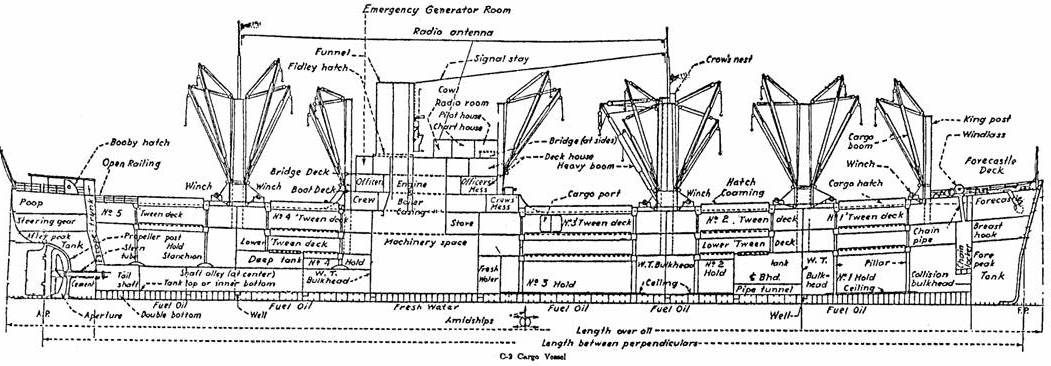 Fig.3
Fig.3
Radiodignosis respiratory system. Radiological symptoms of the respiratory organs diseases
X-ray diagnosis.
Methods of X-ray diagnosis of respiratoty system are divided on: methods of X-ray diagnosis using contrast agents, methods of X-ray diagnosis without contrast agents and radiofunctional methods of X-ray diagnosis.
To the methods of X-ray diagnosis without contrast agents, there are: fluorography, X-radiography, photofluoroscopy, tomography, computer tomography, electro-sciagraphy
Methods of X-ray diagnosis using contrast agents, there are: bronchography, angiopulmonography, artificial pneumothorax, pleurography, pneumomediastinography, fistulography and others.
Functional methods of X-ray diagnosis, there are methods which use X-ray diagnosis in the different respiratory phase.
The fluorography (see fig 10.13) is applied for prophylactic examination of the population.
The roentgenoscopy is used for examination of respiratory organs in more cases(see fig 10.11).
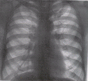
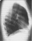
Fig.14.1 Sciagrams of normal thorax are in anterior view and lateral projection
The linear tomography (see fig 10.14, 10.20 b) is used for examination of structure of the pathological formations into a trachea, a large bronches, a root of lung and mediastinum.
Computer tomography (see fig 10.20 c) is used for examination of structure of the pathological process and for estimation the condition of adjacent organs. It is possible to distinguish shades of the pulmonary vessels, the bronches and lymphatic nodes in thoracic cavity. The pulmonary tissue has -650 – -850 НU, mediastinal adipose tissue - 70 – - 120 НU.
Spiral CT-angiography – it’s method of the lungs vessels defeats diagnosis. It gives diagnostic information about cause of narrowing|nature|, morphology of atherosclerotic plaque and vascular wall, about pathological arteries’ deformation. The method allows to find the location of vessels thrombus.
The magnetical resonance imaging is used after computer tomography mainly for evaluation of condition of the mediastinum, because pulmonary tissue gives a weak signal.
The bronchography (see fig 15.2) was used for estimation the trachea and bronchi morphological changes, congenital abnormality, bronchoectasis, bronchial fistules and tumours with the contrast agents which were entered through a catheter.
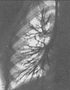

Fig.15.2 a) normal bronchogram of the right lung, anterior view: b) lateral bronchogram of a patient with bronchoectatic disease.
The angiopulmonography (see fig 15.3) – lungs’ vessels contrast examination after catheterisation of the femoral vein (general angiopulmonography). For selective angiopulmonography catheter is entered trought subclavian vein to the necessary pulmonary, lobar or segmentar artery under X-ray control. Indication for angiopulmonography is a suspicion on thrombembolia of pulmonary artery and congenital abnormality of lungs vessels, such as arteriovenous fistulas and aneurysm.
Sometimes contrast agency is injected into the bronchial arteries for differential diagnosis between the lungs tumours|swelling| and the chronic inflammations of lung.
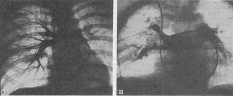
Fig.15.3 Angiopulmonograma
The ultrasound (see fig 15.4) is used for evaluation of condition of the: heart, mediastinum, pleura, diaphragm, superficial parts of lungs. Ultrasonography allowes to find of liquid into a pleural cavity and estimate integrity of ribs. Ultrasound does not penetrate into a deep pulmonary parenchym.
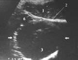
Fig.15.4 The malignant tumour in lungs: 1 liver: 2 diaphragma; 3 tumour; 6 diafragmal pleura
The needle biopsy (see fig 15.5)– it is excision of the lungs tissues under roentgencontrol with bronchoscope through nasal ways (aspirational bronchoscopy), transbronchial biopsy (puncture biopsy through a bronchial wall) or a puncture biopsy through a thoracic wall.
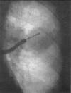
Fig.15.5 A transbronchial biopsy of the left lung upper lobar bronchus
Date: 2014-12-28; view: 4053
| <== previous page | | | next page ==> |
| PILLARS AND GIRDERS | | | The thoracic organs roentgenanatomy |