CATEGORIES:
BiologyChemistryConstructionCultureEcologyEconomyElectronicsFinanceGeographyHistoryInformaticsLawMathematicsMechanicsMedicineOtherPedagogyPhilosophyPhysicsPolicyPsychologySociologySportTourism
Diagnostics
A well-conducted physical examination is enough to confirm the diagnosis of acute leg ischemia, determine the level of obstruction, and evaluate the severity of ischemia. When the leg is immediately threatened, further radiologic examinations or vascular laboratory tests should not under any circumstances delay surgical treatment. When the extremity is viable or marginally threatened, angiography should be performed. Duplex ultrasound is of limited value for evaluating acute leg ischemia and angiography is recommended for almost all patients in these two groups. If angiography is not available or if examination of the patient has verified that emboli is the cause and probably is best treated by embolectomy, angiography can be omitted. This situation is rare, however. The arteriogram provides an anatomical map of the vascular bed and is very helpful in discriminating embolus and thrombosis. The former is essential for planning the surgical procedure, and the latter may be of importance for selecting the treatment strategy.
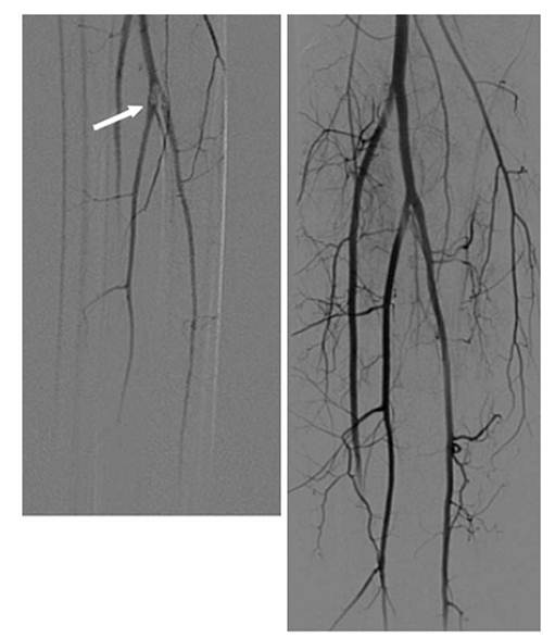
An arteriogram representing an embolus is shown in Fig. 3.
Fig. 3. Embolus lodged at the origins of the calf vessels (arrow). Angiograms display ilms before and after thrombolysis
Angiographic signs of embolism are an abrupt, convex start of the occlusion and lack of collaterals. Thrombosis is likely when the arteriogram shows well-developed collaterals and atherosclerotic changes in other vascular segments. For most patients with viable and marginally threatened legs the diagnostic angiography is followed by therapeutic thrombolysis right away.
Angiography can be performed during daytime when qualified radiology staff is available. The patient should be optimized according to the recommendations given in the next section. Before angiography it is important to keep the patient well hydrated and to stop administration of metformin to reduce the risk of renal failure. Disturbances in coagulation parameters may interfere with arterial puncture and must also be checked before the investigation. The information is also important as baseline values in case of later thrombolysis. The groin of the contralateral leg is the preferred puncture site for diagnostic angiography. A second antegrade puncture can be done in the ischemic extremity if thrombolysis is feasible.
Date: 2014-12-29; view: 890
|