CATEGORIES:
BiologyChemistryConstructionCultureEcologyEconomyElectronicsFinanceGeographyHistoryInformaticsLawMathematicsMechanicsMedicineOtherPedagogyPhilosophyPhysicsPolicyPsychologySociologySportTourism
Reproduction and development
Introduction
A child is born. Life is passed from one generation to the next. By the time of birth, an infant has already developed for nine months within its mother. During these nine months, a single fertilized cell has become a complete human being. From the time of conception to the time of birth, the individual has increased in size 2 billion times! It has developed arms, legs, and intemai organs to carry out all of life's functions. The development of a human being is a common, but very extraordinary, occurrence. Today, worldwide, about 141 children are born each minute, 8,500 each hour, and 74 million each year.
Like all mammals, humans reproduce sexually. Special structures in the male and female make up the human reproductive systems.
The Reproductive Systems
The reproductive organs in males and females are called go-nads, Gonads are present at birth. They develop fully during puberty, a period during which boys and girls mature physically and sexually. In males puberty generally occurs between the ages of 13 and 15. In females puberty is usually complete by 13 years of age. Following puberty gonads produce sex ceils called gametes.
The Male Reproductive System
The male gonads, the testes, are a pair of organs with two primary functions: toproduce male gametes, called sperm, and to produce male hormones. The testes are suspended below the abdomen in an external sac called the scrotum, where the temperature is lower than normal body temperature. The lower temperature is necessary for the production of healthy sperm. Inside the testes are hundreds of tightly packed, coiled tubes, caiied seminiferous tubules, where sperm are produced.
Sperm are carried from the seminiferous tubules to a coiled tube, the epididymis. From the epididymis sperm move into the vas deferens, lightly coiled tubules where sperm are stored. Muscular contractions, called ejaculation, force sperm from the vas deferens through the urethra, in the penis, in the outside of the body.
As sperm are ejaculated, they are mixed with secretions from several glands—the seminal vesicles, Cowper's glunds, and the prostaie. The secretions contain nutrients, hormones, and enzymes thai provide the sperm with energy and the proper environment. The sperm and [he secretions together form a thick, milky liquid called semen.
A human sperm cell is extremely tiny. Approximately 4 billion sperm could easily fit into a thimble. Each sperm cell has three parts—the head, the middle region, and the tail. The head is the cell nucleus. A small region at the tip of the head, the acrosome, contains enzymes that enable the sperm to penetrate the female gamete. The middle region contains the mitochondria that supply the cell's energy. The tail, a long, slender flagellum, uses that energy to move the sperm cell.
During puberty, cells surrounding the seminiferous tubules begin to secrete male hormones, or androgens, the most important of which is lestosterone. Testosterone stimulates development of secondary male sex characteristics. Among these are strong muscles, a deep voice, and body hair. Two tropic hormones secreted by the anterior pituitary are necessary for normal functioning of the testes. LH (luteinizing hormone) stimulates the production of testosterone. FSH (follicle stimulating hormone) controls the maturation of sperm cells.
The Female Reproductive System
The female gonads. the ovaries, are located in the lower pan of the abdominal cavity. These olive-sized organs produce the female gametes, called am or eggs. Below the ovaries is the uterus, a hoilow. thick-walled, muscular organ about the size of a fist. The uterine walls are lined with mucous membrane that contains many glands and blood vessels. An unborn child develops here. A duct, caiied a Fallopian tube, or oviduct, extends from each side of the titerus. Fringed projections at the upper end of each Fallopian tube surround each ovary. Cilia lining these projections propel the mature egg to the uterus. A tube called ihe vagina leads from the uterus to the outside of the body. Between ihe uterus and the vagina is a muscular ring, the cervix.
At birth a female has about 2 million immature eggs in her ovaries. Beginning at puberty a single egg matures and is released from an ovary each month. In general, the ovaries alternate in releasing eggs. The left ovary releases an egg one month, and the right ovary releases one the next month. Approximately 400 eggs mature during a female's lifetime. During pubeny the ovaries aiso begin to secrete ihe female hormone estrogen. Estrogen causes development of secondary female sex characteristics, such as wide hips, body hair, and enlarged breasts.
The Menstrual Cycle
The female reproductive system has three primary functions: the production of eggs, the secretion of female sex hormones, and the nourishment and protection of a new individual. Approximately every 28 days, one egg matures and is released from the ovary, and the uterus is prepared to receive it. These activities are controlled by hormones operating on a feedback system. The entire process of ovuiation and related changes in the uterus operates on a regular, repeating pattern called the menstrual cycle.
The cycle starts with the release of FSH and LH from the anterior pituitary. These hormones trigger the maturing of an egg and its follicle, the fluid-filled chamber around the egg. The follicle secretes estrogen, which causes the uterine lining to thicken. Estrogen also triggers an increase in LH. The LH causes the follicle to rupture and release the mature egg from the ovary surface. The process of releasing a mature egg from the ovary is called mutation.
Following ovuiation LH stimulates the follicle to enlarge and fill with blood vessels. The follicle is now called the corpus luteum, or "'yellow body." It functions temporarily as an endocrine gland, producing the ovarian hormones estrogen and progesterone. Progesterone continues the process of thickening the uterine lining in preparation for the uterus to receive a fertilized egg. Both estrogen and progesterone inhibit the production of GnRH (gonadotropin-releasing hormone) by the hypothalamus. The lower GnRH level inhibits the production of FSH and LH, thus keeping another follicle from maturing.
If an egg is not fertilized, the corpus luteum disintegrates. The lining of the uterus collapses, (is cells die and are sloughed off through the vagina. This discharge of dead tissue and the unfertilized egg is called menstruation. The decreased levels of FSH and LH, plus disintegration of the corpus luteum. lead to lower progesterone and estrogen levels. The Eower hormone levels trigger GnRH, which stimulates FSH and LH secretion. Thus a new cycle begins.
Males normally produce healthy sperm until about age 80. Females, however, cease to produce mature egg cells afier menopause, the time at which the menstrual cycle ceases. This period usually begins between the ages 45 and 55. After menopause the pituitary does not secrete LH and follicles do not mature. Therefore, estrogen and progesterone are not produced.
Fetal Development and Birth
The fusing of a sperm nucleus and an egg nucleus is called fertilization. When an egg is fertilized, menstruation does not occur. Instead, a nine-month gestation period begins. This period, also called pregnancy, is the time during which a fertilized egg develops into a child inside the mother.
Fertilization
During sexual intercourse semen is ejaculated through the male's penis into the female's vagina. Of some 400 million sperm ejaculated, less than 3,000 sperm get as far as a Fallopian tube and, of these, only about 50 reach the mature egg. In fertilization the head and middle section of the sperm enter the ovum. Only one sperm can fertilize an egg. Changes occur in the egg cell membrane to prevent other sperm from penetrating it.
Since both gametes are haploid, each parent contributes half the normal chromosome number. In human beings the haploid number is 23 chromosomes. When the gametes fuse, the zygote, or fertilized egg, has 46 chromosomes.
Thinking About Biology: Hope for the Childless
Blockage of the Fallopian tubes is the chief reason why many women are unable to become pregnant. The blockage prevents sperm from entering the tube and fertilizing a mature egg.
The technique known as in vitro fertilization (IVF) can overcome this difficulty in some cases. The term in vitro means "in glass" and refers to the fact that fertilization takes place in a laboratory dish rather than in the human body.
The procedure involves taking a mature egg from the woman's body when the egg is ready to be released from the ovary.
A physician removes the egg through a small incision in the woman's abdomen. The egg is placed in a laboratory dish to which nutrient has been added to keep the egg healthy. Sperm from the husband is added to the dish. After two days the material in the dish is examined to determine whether the fertilized egg is dividing. If division is taking place, the egg is implanted in the woman's uterus, ff the implantation is successful, the woman becomes pregnant. Nine months later, the newborn infant joins the ranks of "test tube" babies.
In vitro fertilization has made ft possible for a number of childless couples to have babies. However, these successes represent only about 12 percent of the women who undergo IVF. Most failures occur during implantation. The removal of the unfertilized egg from the ovary can cause bleeding or a hormone imbalance. When that happens, it means that the lining of the uterus is not ready to accept the egg at the time of implantation, and pregnancy will not occur.
Embryonic Development
About 36 hours after fertilization, zygote begins to devide by mitosis. The zygote continues to devide, forming a hollow ball of cells called the blastula. At this stage a major transformation occurs. The inner cell mass develops three layers, which eventually develop into the variousorgans of the body. This process is called gastrulation. The blastula then moves down the oviduct and into the uterus where it is now called a blastocyst. Only one part of the blastocyst will develop into embryo. An unborn child is called an embryo during the first eight weeks of its development. Afterward it is referred to as a fetus. The outer blastocyst cells, called the trophoblast, are not part of the embryo.
Six days after fertilization, the blastocyst attaches itself to the lining of the uterus, a process called implantation. From the trophoblast cells, four protective membranes are formed. They are the yolk sac, allantois, amnion, and chorion. The yolk sac has no yoik, as it does in birds, and serves no major function in the human embryo. The allantois functions in the formation of fetal blood vessels and gives rise to the umbilical cord. The umbilical cord, which contains two arteries and one vein, is the chief connection with the mother's uterus and permits the embryo to float in fluid. The amnion forms a sac, which fills with fluid. This amniotk fluid cushions the embryo and keeps it moist. The outermost membrane, the chorion, forms fingerlike projections, called villi, that attach to the lining of the uterus. Blood vessels from the ailantois run through the villi.
Together the chorion and allantois form the placenta. The placenta is a mass of tissue that secretes estrogen and progesterone, which help maintain a thickened, blood-enriched uterine lining. The placenta also serves as the point of exchange of substances between mother and fetus. The exchange takes place through the umbilical cord by which the fetus is attached to the placenta. However, the connection between the circulatory systems of the mother and child is not a direct one. The mother's body carries out the processes of digestion, excretion, and respiration for the developing child. Food and oxygen pass from the mother's bloodstream into the capillaries within the villi of The placenta. The food and oxygen then pass into adjacent capillaries belonging to the fetus and, thus, into the main bloodstream of the fetus. Waste products pass from the bloom If the fetus to the blood of the mother in the same manner.
The nine-month pregnancy is commonly divided into three three-month periods called trimesters. During the first trimester, unorganized cells become organized into vital organs. In the second trimester, the organs and body features become more refined. After 25 weeks, development is complete. During the final trimester, most growth in body size takes piace. A chiid born at this time would be premature but. with proper care, it would have a good chance of surviving outside its mother's body.
Childbirth
After approximately 266 days inside its mother's body, the child is ready for birth. Shortly before birth the mother's body undergoes changes that prepare it for labor, the process of literally forcing the child our of her body. Contractions of uterine muscles become progressively stronger due to a decrease in progesterone, an increase in estrogen, and the release of the hormone oxytocin by the hypothalamus. Oxytocin speeds up contractions of the uterus. The cervix relaxes and widens from about 0.5 cm to 10 cm. As the baby enters the birth canal, the amnion breaks, and amniotic fluid escapes. Muscle contractions become very strong and finally push the infant out of the mother's body. The passing of the child from the uterus into the external environment is called delivery.
After delivery the umbilical cord is cut and tied to prevent a loss of blood through the umbilical blood vessels. The child must now support its own life processes. During the gestation period, fetal lungs do not operate. At birth carbon dioxide from cellular activity quickly builds up in the child's blood. This buildup triggers the respiratory center in the brain to take in oxygen, and the child begins breathing on its own.
Fetal blood follows a path that bypasses the lungs. Freshly oxygenated blood flows from the placenta to the heart and deox-ygenated fetai blood flows through the heart in a path which leads it back to the placenta where it is oxygenated once again. Two openings in the fetal heart make this circulation possible.
Human hyealth and environment
People’s wellbeing and health have always been depended on the quality of environment, but the last decade the environmental situation has become so complicated and insecure that these issues became very urgent and attract very serious attention.
When we speak about health we imagine an ordinary, typical state of functioning of an organism or a system. When we say “healthy person” we mean a concrete person who doesn’t have any diversions in physiological and psychological spheres. When we say “healthy nation” we presuppose that indicators of health of every representative of this nation in total have quite a satisfactory level. When we say “healthy economy” we mean that we speak about a typical economic system that is developed according to ordinary rules and which doesn’t seem to cause any worries about its unpredictability in the future. In the latter case the notion of “health” acquires a bit different meaning as it goes beyond medical meaning. But whenever we use the notion of health we mean the normality of development of anything we talk about.
So, health is a state of an organism (or in a wider sense of a system) when all its organs (system components) are able to function according to the normal processes.
Human health is an integral notion and is characterised by parameters which are related to its physical, psychological and social essence.
Numerous observations and special research demonstrate that human health is very much dependant on lots of factors of natural development (figure 6.1.)
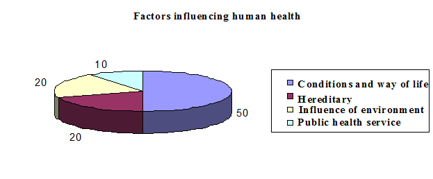
Figure 6.1. Conditions influencing human health
All known factors of the environment can influence the state and development of the organisms and that is why at the end of the day they predetermine qualitative characteristics of populations. The majority of the factors are necessary for the development of living organisms; though, there are also such factors that have a destructive influence on them. In the table below we demonstrate the examples of the influence of some environmental factors on human health.
Table 6.3.
Influence of external environmental factors on human health
| № | Environmental factors | Influence on human organism |
| Thermal field, temperature of the environment | Under deviation from optimal temperature (20–22 оС) to the increase or decrease metabolism is changing, organ’s and system’s biorhythms are disrupted and inflammatory and allergic reactions are aggravated. It is believed that thermal regime of the environment influences morphometric and anatomic indicators of the human’s organism. | |
| Atmospheric pressure | Change of atmospheric pressure causes disruption of metabolic processes and can initiate hyper and hypotonic crises, headaches and decrease ability to work and increase intensification of chronic diseases. | |
| Humidity | Excessive humidity facilitates activation of respiratory and inflammatory processes and assists in formation of the centres of epidemic diseases. | |
| Chemical components of the environment | They have a wide range of influence on the human body – from stimulation of some functions of growth and development to suppression and death. | |
| Gravitation | Change of intensity of gravitation (under artificial conditions or, for example, at different Moon phases) influences at least on irritability of nervous system and other functions of the organism. | |
| Magnetic and electromagnetic fields (magnetic storm, local magnetic fields of domestic appliances and so on) | Change of intensity and frequency of vibrations of electromagnetic and magnetic fields cause hypertonic and hypotonic crises, facilitate stenocardia, disrupt heart rhythm, increase the quantity of heart attacks and diseases, increase capillary pressure, change nervous, endocrine and immune system, as well as influence red-ox processes at cellular and sub-cellular level. Other deviations are also possible. | |
| Noise and vibrations | They decrease level of attention, deteriorate general condition, increase the quantity of mistakes, cause stress and oppress central neural system, provoke change of pulse and breath level, disrupt metabolism and facilitate hypertension and heart diseases. | |
| Ionising radiation | It could provoke radiation sickness, malignant tumour, leucosis, and induce intensification of hereditary diseases. Marrow, small intestine and central nervous system are most sensitive to ionising radiation. |
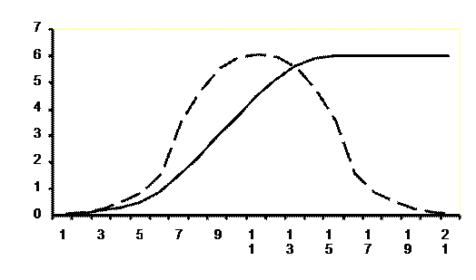
It is a given general principle of the influence of ecological factors on human’s and animal’s organisms that the effect or intensiveness of its influence depends on the nature of the factor itself and on its quantity (for chemical factors) or dose (for physical factors). Those dependencies of the influence of concentration or dose of the certain ecological factor on this or that vital indicator (in the long run it could be the quantity of population) are non monotonous and are generalised on the figure 6.2.
  |
 |
 |
Figure 6.2. Graphic demonstration of generalised dependence of intensive influence of ecological factors (dose, conventional units) on vital indicators of the organism or population (effect, conventional units)
Interrelation between the state of the environment and people’s health has become unique and indisputable. High quality of the environment does not guarantee 100% health of the population but its influence would not become aggravating for the people’s health. At the same time deterioration of the environment resulting from human activity inevitably increases risks for human health. It should be known and remembered, at the least. At the most, people’s life strategies should be designed in such a way so that it minimises negative impact on the environment, the environment, which we are living in.
Date: 2014-12-22; view: 1413
| <== previous page | | | next page ==> |
| Vitamins and Minerals | | | World populationand its regulation |