CATEGORIES:
BiologyChemistryConstructionCultureEcologyEconomyElectronicsFinanceGeographyHistoryInformaticsLawMathematicsMechanicsMedicineOtherPedagogyPhilosophyPhysicsPolicyPsychologySociologySportTourism
Histologic and Cytologic Methods.
The laboratory diagnosis of cancer is, in most instances, not difficult. The two ends of the benign-malignant spectrum pose no problems; however, in the middle lies a gray zone where one
should tread cautiously. The focus here is on the roles of the clinician (often a surgeon) and the pathologist in facilitating the correct diagnosis.
Clinical data are invaluable for optimal pathologic diagnosis, but often clinicians tend to underestimate the value of the clinical data. Radiation changes in the skin or mucosa can be similar to those associated with cancer. Sections taken from a healing fracture can mimic an osteosarcoma. Moreover the laboratory evaluation of a lesion can be only as good as the specimen made available for examination. It must be adequate, representative, and properly preserved. Several sampling approaches are available: (1) excision or biopsy, (2) needle aspiration, and (3) cytologic smears. When excision of a small lesion is not possible, selection of an appropriate site for biopsy of a large mass requires awareness that the margins may not be representative and the center largely necrotic. Appropriate preservation of the specimen is obvious, yet it involves such actions as prompt immersion in a usual fixative (commonly formalin solution, but other fluids can be used), preservation of a portion in a special fixative (e.g., glutaraldehyde) for electron microscopy, or prompt refrigeration to permit optimal hormone, receptor, or other types of molecular analysis. Requesting "quick-frozen section" diagnosis is sometimes desirable, for example, in determining the nature of a mass lesion or in evaluating the margins of an excised cancer to ascertain that the entire neoplasm has been removed. This method permits histologic evaluation within minutes. In experienced, competent hands, frozen-section diagnosis is highly accurate, but there are particular instances in which the better histologic detail provided by the more time-consuming routine methods is needed—for example, when extremely radical surgery, such as the amputation of an extremity, may be indicated. Better to wait a day or two despite the drawbacks, than to perform inadequate or unnecessary surgery.
Fine-needle aspiration of tumors is another approach that is widely used. The procedure involves aspirating cells and attendant fluid with a small-bore needle, followed by cytologic examination of the stained smear. This method is used most commonly for the assessment of readily palpable lesions in sites such as the breast, thyroid, and lymph nodes. Modern imaging techniques enable the method to be extended to lesions in deep-seated structures, such as pelvic lymph nodes and pancreas. Fine-needle aspiration is less invasive and more rapidly performed than are needle biopsies. In experienced hands, it is an extremely reliable, rapid, and useful technique. Cytologic (Pap) smears provide yet another method for the detection of cancer ( Chapter 22 ). This approach is widely used development of tests to detect cancer markers in blood and body fluids is an active area of research. Some of the markers being evaluated include the detection of mutated APC, p53, and RAS in the stool of patients with colorectal carcinomas; the presence of mutated p53 and of hypermethylated genes in the sputum of patients with lung cancer and in the saliva of patients with head and neck cancers; and the detection of mutated p53 in the urine of patients with bladder cancer.[207]

Figure 7-54 A normal cervicovaginal smear shows large, flattened squamous cells and groups of metaplastic cells; interspersed are some neutrophils. There are no malignant cells. (Courtesy of Dr. P.K. Gupta, University of Pennsylvania, Philadelphia, PA.)

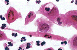
Figure 7-55An abnormal cervicovaginal smear shows numerous malignant cells that have pleomorphic, hyperchromatic nuclei; interspersed are some normal polymorphonuclear leukocytes. (Courtesy of Dr. P.K. Gupta, University of Pennsylvania, Philadelphia, PA.)

Figure 7-56Anticytokeratin immunoperoxidase stain of a tumor of epithelial origin (carcinoma). (Courtesy of Dr. Melissa Upton, University of Washington, Seattle, WA.)
TABLE 7-13-- Selected Tumor Markers
Date: 2016-04-22; view: 1084
| <== previous page | | | next page ==> |
| GRADING AND STAGING OF TUMORS | | | Markers Associated Cancers |