CATEGORIES:
BiologyChemistryConstructionCultureEcologyEconomyElectronicsFinanceGeographyHistoryInformaticsLawMathematicsMechanicsMedicineOtherPedagogyPhilosophyPhysicsPolicyPsychologySociologySportTourism
LABORATORY DIAGNOSTIC OF THE OPORTUNISTIC INFECTIONSActual theme.The problem of theopportunistic infections is very important today. In the last year different pus-inflammatory disease are registered often. Its causative agents are opportunistic infections. It is necessary to detail study etiology, pathogenesis, clinic and diagnostic of the opportunistic infections because there are many different causative agents (bacteria, viruses, fungi, protozoa) of this diseases, different clinical manifestation, the lesion humans with different age, sоcial status, education. Primary objectives:to be ableto conduct and evaluate the microbiological diagnostics of the opportunistic infections. The principles of the diagnostic, treatment and prevention this infections are. QUESTIONS FOR DISCUSSION 1. The exogenous opportunistic infections: pseudotuberculosis, serraciosis, pseudomonios at all. 2. Еndogenous opportunistic infections, the importance of the members of the normal flora in its arising. 3. Microbiological diagnosis of opportunistic infections. The etiological criterion of the meaning of the microorganisms that isolated from center of the pathology. 4. Etiologic, pathogenesis, diagnostic and principles of prophylaxis and medical treatment of the widely spread opportunistic infections: - Bacteriemia and sepsis (labor, wound, urogenital, surgery at all.) - pus-inflammatory infections: wound, burn, postnatal, postoperative, peritonitis, оsteomyelitis at all) - bronchopulmonary infections: acute and chronic bronchitis, acute and chronic pneumonia, pulmonary abscess. - Urological infections: pyelonephritis, cystitis, urethritis, prostatitis. - acute intestinal infections: food poison and food intoxications; - Infections of CNS: meningitis, encephalitis, abscess; - мycobacteriosis; - мycoses. PROCEDURE OF PRACTICAL SESSION Task 1. Study the smears from the patient’s material. Task 2. Study the growth of Staphylococcus aureus, Streptococcus pyogenes, Klebsiellla pneumoniae, Pseudomonas aeruginosae, Escherichia coli. RECOMMENDATIONS FOR PRACTICAL WORK Task 1.
In the smear from patient with infection caused by Pseudomonas aeruginosae, we see Gram-negative rods that are one by one.
In the smear stained by Burry-Ginze from patiens with pneumonia caused by Klebsiellla pneumoniae discoloring capsule and pink cytoplasm can be seen.
Task 2.
Staphylococcus on the EYA forms middle, convex, opaque, homogeneous, gold, white or yellow colony. On the EYA growth another bacteria is inhibiting. A ring of the dimness forms a round of the Staphylococcus colony because it is producing enzyme lecitinase.
Salmonella on the Endo forms circum, convex, semi-transparent pink colony because it is not producing enzyme lactose.
Klebsiella pneumoniaeforms on Ploskirev’s medium Lac positive colonies.
Addition 1 Date: 2016-01-14; view: 950
|
 In the smear from the pus Staphylococcus are circum violet microorganisms placed in accumulation as bunch of the grapes.
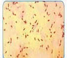
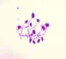 In the smear from patients with candidiasis big oval Gram positive microorganisms are observed, in some fields there are mycelium and budding cells.


 Pseudomonas aeruginosa on the MPA forms circum, dry or smooth (in capsule forming bacteria) colony. The culture of the Pseudomonas aeruginosa have specific smell, like a jasmine or strawberry soap.