CATEGORIES:
BiologyChemistryConstructionCultureEcologyEconomyElectronicsFinanceGeographyHistoryInformaticsLawMathematicsMechanicsMedicineOtherPedagogyPhilosophyPhysicsPolicyPsychologySociologySportTourism
RECOMMENDATIONS FOR PRACTICAL WORK
 Task 1.
|
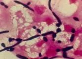
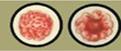
 The material for research was staining by Gram method. The cells of Candida spp. appear Gram-positive with a blue-black appearance. This micrograph illustrates the dimorphic nature of this fungus with yeast cells and mycelia both being present. Mycelial cells are more commonly found in cases of clinically apparent candidosis. Candida spp. will be yeast-like with an asympomatic infection.
Task 2.
|


|
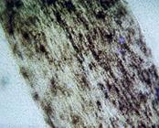
 In the smear stained by Gram method there are large Gram-positive cells arranging like butterfly wings.
Task 3.
|
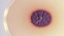
 Trichphiton rubrumand Trichophyton violaceum can be cultivated on the Saburo agar. Material before inoculation is processing by antibiotics and incubating by temperature 30°C. In positive cases, growth is observed on 3 - 5 days as various colonies. Trichophyton rubrum forms colonies with a red pigment. This feature is characteristic only for this type of fungus.
|
Task 4.
At determining the amount of the fungus it is needed to take into account in 1 ml of sputum, that a sputum was preliminary divorced in 100 times, and on Petri dish sowed 0.1 ml of the divorced sputum. It is needed to count up the amount of colonies of fungus which grow on Petri dish and to increase the got number on 1000 (taking into account aforesaid). If amount of fungus ≥ 100 000/ml material, it grounds to diagnose «Candidiasis». Amount of fungus from 50 000 to 100 000/ml material is an index higher than norm and for clarification of diagnosis, in this case, it is needed to conduct the repeated research in a few days. The colonies of fungus selected in an insignificant on Petri dish are estimated as carrier.
Task 5.
Polysacharade antigen from Candida spp. for CFTis extract of polysacharade antigens of Candida spp., used for diagnostics of Candidiasis in serological method (for determination of specific antibodies in the serum of blood of patient). A reaction (CFT) is conducted with pair serum.
Erythrocytes Candida diagnosticum conteins the antigens of Candida spp. that are adsorbed on the erythrocytes. This diagnosticum used for serological diagnostic of Candidiasis in PHAT with pair serum.
Task 6.
The concentration of antibodies in the serum of the patient with Candidiasis does not arrive at high titles, that is why it is important to trace the changes of their concentration during a disease. A diagnostic value has a fact of increase of titre of anticandidiasis antibodies in 4 times and anymore. A positive reaction is characterized formation of grainy sediment of large-break with an unequal edge as an “umbrella”, while in small holes, where a reaction is negative; there is compact sediment with an even edge as a “button”.
The components of PHAT are: NaCl-solution, pair serum from patient (unknown antibody), erythrocytes Candida diagnosticum (known antigen). Because fungus of Candida are a normal microflora a serum reaction needs to be put in a dynamics. Diagnosis Candidiasis is put on increase of titer of antibodies in a pair serum. For example:


| 1:20 | 1:40 | 1:80 | 1:160 | Control of the serum | Control of the antigen |
Addition 1
Date: 2016-01-14; view: 3156
| <== previous page | | | next page ==> |
| Laboratory diagnostics of Mycoplasma and Chlamidia diseases | | | Laboratory diagnostics of mycosis |