CATEGORIES:
BiologyChemistryConstructionCultureEcologyEconomyElectronicsFinanceGeographyHistoryInformaticsLawMathematicsMechanicsMedicineOtherPedagogyPhilosophyPhysicsPolicyPsychologySociologySportTourism
PROCEDURE OF PRACTICAL SESSION
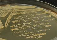
Task 1. Study the preparation from the pure cultures of Staphylococcus areus and S.epidermidis microscopically; draw them in the protocol.
Task 2. Prepare and stain by Gram method the preparation taken from the blood of the patient suspected of staphylococcal sepsis, draw it in the protocol.
Task 3. Study the growth of S. aureus and S. epidermidis in milk meat infusion agar. Fill in the protocol with their cultural properties.
Task 4. Study the growth of S. aureus and S. Epidermidis in blood meat infusion agar. Fill in the protocol with their cultural properties.
Task 5. Study the growth of S. aureus and S. Saprophiticus in egg yolk salt agar. Fill in the protocol with their cultural properties.
Task 6. Study the growth of other types of Staphylococcus spp. in citrate plasma. Draw the plasma in the test tubes before streaking of Staphylococcus spp. and after 24-hour growth.
Task 7. Study the sensitivity of the isolated staphylococcal culture to antibiotics; draw a conclusion. Write the results in the protocol.
Task 8. Study the sensitivity of the isolated staphylococcal culture to phintonside garlic and make a conclusion. Write the results in the protocol.
Task 9. Study the main antimicrobial drugs used for treatment, prevention and diagnostics of suppurative diseases. Write them in your copybook.
RECOMMENDATIONS FOR PRACTICAL WORK
Task 1.
 All types of Staphylococcus spp. have identical staining and morphological properties. Therefore, stained by Gram method of pure culture of S.aureus, S.epidermidis in preparation it is possible to see gram-positive cocci like "clusters of vine".
Task 2.


| |
| |
| |
|
Task 3.


Milk meat infusion agar is a special medium capable to expose the ability of bacteria to form a pigment. Therefore, S.aureus forms yellow colonies in this medium. That is why they are called golden staphylococci. Other types of staphylococci do not have such a pigment.
Task 4.
Blood agar is a special medium with the ability of bacteria to select hemolysin coming to light on. In blood agar abundant growth of the most staphylococcal species occurs within 18 - 24 hours. Only individual colonies should be picked for preliminary identification testing at this time. Since most species can not be distiguished from each other in the basis of the colony morphology within a 24-hour incubation period, colonies should be allowed to grow for at least additional 2 - 3 days before the primary isolation plate is confirmed for species or strain composition.
|
|


|


|

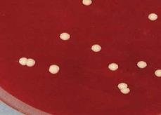
Task 5.
Task 5
 For revealing lecitinase activity of Staphylococcus spp., cultures of bacteria grow on the special selective medium. This medium is yolk salt agar containing 8-10% of NaCl. Staphylococcus spp. tolerates to sodium chloride in concentrations of 5-10%. Salt-containing media are useful in isolating staphylococci from samples containing large numbers of other bacteria. The bacteria allocating lecitinase destroy yolk lecithin. Therefore round colonies with the turbidity zone (as enzyme diffusion on agar) are formed. Such bacterium in this case is S.aureus.
Other kinds of staphylococci, such as S.epidermidis, S.saprophyticus, do not allocate such enzyme, therefore round colonies do not form lecitovitelase zone.
Task 6.

|
|
Task 7.

Measure a zone of absence of growth round a paper disk, study the sensitivity of isolated staphylococcal culture to antibiotic, and make the conclusion. Put down the results in the protocol.
Task 8.


Staphylococcus spp. is known to be sensitive to vegetative antibiotics. In the given experience, because of diffusion phytoncide garlic in an agar the growth of inhibition zone is formed. It specifies the bactericidal action of the given vegetative antibiotics on the given culture.
Task 9.
Staphylococcal toxoid is a vaccine containing inactivated, which help formalin (0,4%) and temperature (40°C) exotoxin S.aureus. It is used specifically to prevent and treat staphylococcal infections.
Addition 1
Date: 2016-01-14; view: 3681
| <== previous page | | | next page ==> |
| Microbiological diagnosis of the botulism | | | Laboratory Diagnostics of Staphylococcal Infections |