CATEGORIES:
BiologyChemistryConstructionCultureEcologyEconomyElectronicsFinanceGeographyHistoryInformaticsLawMathematicsMechanicsMedicineOtherPedagogyPhilosophyPhysicsPolicyPsychologySociologySportTourism
The radial methods investigation of the musculoskeletal system. The radio symptoms of pathology of the musculoskeletal system.
Words and Terms to be Remembered
| dimensions mould moulded overall feature length forward perpendicular after perpendicular breadth | depth draught (draft) extreme designed projecting ratio Lloyd's Register registered measurement |
Read the text and answer the following questions.
1. What are the ship's principle dimensions?
2. What is the difference between the overall and extreme dimensions?
3. Do the principle dimensions influence the ship hull shape?
PRINCIPLE DIMENSIONS OF A SHIP
The principle dimensions of ships are the combination of constructive, designed and overall ship's measurements. They are: length, breadth and draught.
Length between perpendiculars (Lbp) is the distance between fore and aft perpendiculars on the constructive waterline plane. Forward perpendicular (F.P.) is a vertical line through the intersection of the load waterline with the fore side of the stem. After perpendicular (A.P.) is a vertical line through the intersection of the load waterline with the after side of rudder post.
Length overall (Loa) is the greatest length of a vessel from the extreme fore end to the extreme after end without projecting parts. Lloyd's or registered length is measured from the fore edge of stem to the after edge of sternpost at the level of the upper deck.
Breadth (B) or beam is the width of a ship. Extreme breadth is the greatest one to the outside of plating. Breadth moulded (Bm) means the greatest breadth of a ship to the outside of frames.
Depth (H) is the vertical distance measured at the middle of the vessel's length from top of keel to top of upper deck at sides, or to the top of the upper deck beam for designed depth. The designed depth depends on the draught and freeboard required.
Draught, draft (T) is the depth of water which a ship requires to float freely. It's the depth of a vessel below the waterline, measured vertically to the lowest part of the hull, propellers or other reference points. Extreme draft is the vertical distance from the designed waterline to the lowest projecting portion of the vessel at any point in a vessel's length.
The principle dimension ratios give the idea of ship hull shape and potential possibilities of ensuring some operational qualities of a vessel. The greater the L/B ratio, the faster the ship; the greater the B/T ratio, the better the ship stability; the greater the H/T ratio, the higher the degree of unsinkability.
Lbp
A.P. 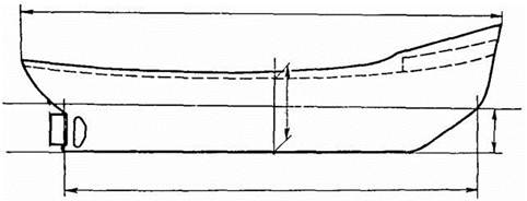 F.P.
F.P.
Fig. 6.
 Exercises and assignments
Ex. 1. Give the initial forms of the following nouns and translate them into Russian:
length, breadth, depth, width, measurements, combination, construction, plating, possibility, operation, reference, unsinkability.
Ex. 2. For the words in (a) find the synonyms given in (b):
a) length, breadth, draught, dimensions, designed, overall, upper, project, ratio, fast, principle;
b) relationship, distance, immersion, width, whole, moulded, measurements, top, high-speed, protrude, basic.
Ex. 3. Give the forms with -ed in the function of attribute from the following verbs. Find corresponding nouns for them in the text and translate the word combinations into Russian:
to design, to measure, to register, to mould, to frame, to require, to receive, to calculate.
Ex.4. Give Russian equivalents for the following word combinations. Consult a maritime dictionary:
moulded dimensions, limiting dimensions, overall dimensions, linear dimensions;
registered length, waterline length, overall length;
extreme breadth, overall breadth;
freeboard depth, registered depth, moulded depth;
after draught, allowable draught, forward draught, full draught, ballast draught, designed draught.
Ex.5. Compare the principle particulars of the two different vessels.
M/V "FORTUNA COAST"
Multipurpose vessel
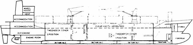
Fig. 7.
| Deadweight Length o.a. Length b.p. Breadth moulded | 1,205 67.35 61.75 11.40 | t; m; m; m; | Depth moulded to main deck Depth moulded to tweendeck Draught Speed | 5.50 3.15 3.51 11.0 | m; m; m; kn. |
M/V "PRESIDENT TRUMAN"
Container carrier
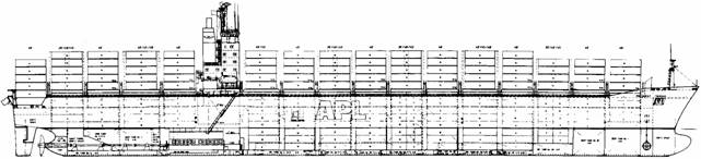
Fig. 8.
| Deadweight Length o.a. Length b.p. Breadth | 54,500 275.20 260.80 39.40 | t; m; m; m; | Depth Max. draught Speed at 11.0 m | 23.60 12.50 24.2 | m; m; kn. |
The radial methods investigation of the musculoskeletal system. The radio symptoms of pathology of the musculoskeletal system.
Section contents:
The roentgenologic methods of the bones and joints visualisation are sciagraphy, tomography, fistulography, pneumoartrography, angiography, and densitometry. The normal roentgenanatomy and bases of physiology of bones and joints, old age features of skeleton structure. The order of study and description of results of roengenological visualisation of bones and joints. Basic indications and contra-indications.
The radionouclear methods of visualisation of bones and joints are planar osteoscintigraphy, SPECT-scintigraphy. The basic principles of radionouclear visualisation of the musculoskeletal system, RPhP, which used for osteostcintigrapgy. The radionouclear semiotics of tumour defect of bones and joints (primary and secondary), inflammatory processes, traumatic damage, degenerative-dystrophic changes of the musculoskeletal system. Basic indications and contra-indications.
The possibilities of US, CT, MRI in visualisation of the musculoskeletal system. Basic indications and contra-indications.
The radiological signs (symptoms) of diseases of the musculoskeletal system are changes of the form, size, position of bones; changes of contours (periosteitis, periostosis, paraostosis), changes of structure (osteoporosis, osteosclerosis, destruction, osteonecrosis, osteolisis and atrophy), changes of joint space (narrowing, disappearance, compression of joint surfaces, regional bone excrescences, disparity of joint ends).
The radiological methods of musculoskeletal system visualisation
1. Roentgenologic method (see fig 11.1): multi-projection (roentgenography) sciagraphy, functional sciagraphy (vertebral column, joints, foot), planar tomography, panoramic tomography (mainly perform to visualisation of jaws). To examine the vessels of musculoskeletal system, as well as other organs and systems, angiography is used. To other X-ray opaque methods belong sinusography (use for examine of paranasal sinuses) and artrography (to purpose for medical survey of joint cavities and used radiolucent (air) or radiopaque agents), fistulography (roentgenography after introduction of radiopaque to the fistula), densitometry (method of estimation of bone tissue density by the agency US or x-ray examination).
1 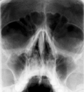 2
2 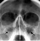 3
3 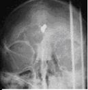
Fig.11.1 X-ray film (sciagram): 1 – maxillary sinuses are norm; 2 – maxillitis; 3 – foreign body into the cerebrum – bullet.
2. The Ultrasound visualisation (US) (see fig11.2) that is sonography of musculoskeletal system is used for the study of soft tissues. It is applying for the examination of children. This method dosn’t have contra-indications. Sonography is more frequently use for estimation of instability of hip joint (at the displasy), the exudate or haematoma in the cavity of large joints, damages of articular cartilage, tendons and ligaments, and also for foreign bodies especially radiolucent. The needle aspiration or biopsy is used under US vision.
1 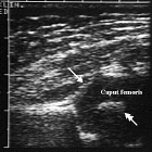 2
2 
Fig.11.2 US: 1 – normal hip joint; 2 - exudates into a joint.
3. The CТ (see fig 11.3) exceeds possibilities of roentgenography; it is performed after previous roentgenologic visualisation. Intravenous contrast for study of location of large vessels in relation to bones and pathological formations is used.

Fig.11.3 The CT: fracture of neck vertebra.
4. The magnetic resonance imaging (MRI) (see fig 11.4) is more advantages CT in the case of visualisation of soft tissues and marrow. Performed in axial, sagital, and coronal (frontal) projections. MRI don’t use for study a bone structure and calcification.
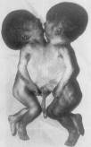
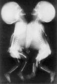
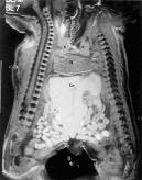
Fig.11.4 Siamese twins: original appearance, sciagrams, MRI.
5.The nuclear imaging of the skeleton visualisation
Indications: visualization of ostiogenic and bone marrow malignant tumours, metastases, benign tumors, definition of activity of inflammatory processes.
Techniques of evaluation:
Therefore by concentration of osteotropnic RPhP in the focal lesions it is possible to estimate intensity of osteogenesis, that allows to differ malignant tumours, non-tumor diseases and inflammatory process. The main osteotropic RPhP are containing the 99mТс, Sr, P and etc.
Scintigraphy of the soft tissues. The method based on selective accumulation of tumourotropnic RPhP in malignant tumours of soft tissues. After intravenous introduction of 300 - 350 Мbк of 99mТс-pertechnat in 0,5 - 2 - 3 hours or in 24 - 72 hours after introduction of 100 - 150 МBк 67Ga-citrate carry out 3 hours static, dynamic or topographical scintigraphy. Levels of accumulation of RPhP in sarcomas of soft tissues is 200 ± 30 %, in benign tumours - 130 ± 20 % (see fig 9.38).
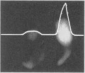
Fig.9.38. Scintigraphy with 99mТс-pirophosphate. Sinovial sarcoma of the right hip. The treefold
(300 %) increase accumulation of RPhP in the focus of lesion.
Osteoscintigraphy, SPECT, PET. The scintigraphy of skeleton is performed in 3—4 hours after intravenous introduction of 300—370 МBк of RPhP. Results of osteoscintigraphy provide qualitative and quantitative estimations. Scintigram distinguish the focal lesions in the sizes from 0, 8-1,0 cm in diameter. In malignant primary and secondary tumours accumulation of RPhP in the pathological focal exceeds up to 150 % (see fig. 9.39.). At inflammatory and degenerate-dystrophic processes accumulation of RPhP does not exceed 150 %.
SPECT and PET are applied in unusual complex cases of diagnostics (see fig. 9.40.)
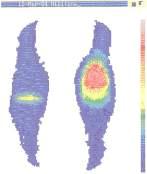
Fig.9.39. Scintigram of knee-joints in frontal view: sarcoma of the left hip.
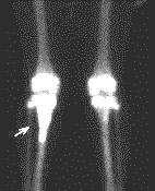
Fig.9.40. PET of knee-joints in frontal view: sarcoma of the right tibia.
Date: 2014-12-28; view: 2167
| <== previous page | | | next page ==> |
| Section 3. Ship’s principle dimensions | | | The radio anatomy of the musculoskeletal system |