CATEGORIES:
BiologyChemistryConstructionCultureEcologyEconomyElectronicsFinanceGeographyHistoryInformaticsLawMathematicsMechanicsMedicineOtherPedagogyPhilosophyPhysicsPolicyPsychologySociologySportTourism
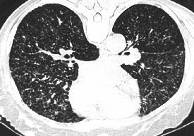
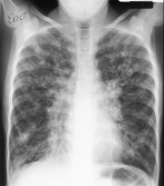
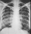
TuberculosisTuberculosis is an infectious disease the exciter of which is microbactery of tuberculosis. Lately in Ukraine there was growth of population morbidity on this disease, which is explained by unfavorable ecological and social terms which influence on reactivity of organism of people, espesially of the so-called marginal elements - drug addicts, alkogolics, persons which are in the places of imprisonment. More frequent an exciter gets to the organism of a man by aerogene way. Clacification of the respiratory organs tuberculosis consists of 14 clinical forms: 1. Primary tubercular complex. 2. Disseminated tuberculosis. 3. Focal tuberculosis. 4. Infiltrative tuberculosis. 5. Caseous pneumonia. 6. Tuberculoma of lungs. 7. Cavernous tuberculosis of lungs, fibrouse-cavernous tuberculosis of lungs. 8. Cirrhotic tuberculosis of lungs. 9. Tubercular pleurisy (including empyema). 10. Tuberculosis of bronches, trachea and superior respiratory tracts. 11. Tuberculosis of respiratory organs, combined with dust professional diseasees of lungs (coniotuberculosis). 12. Outside pulmonary tuberculosis. 13. Tuberculosis of thoraxic lymphatic nodes. 16. Milliar tuberculosis. 16. Other ongans and systems tuberculosis. The clinical forms of tuberculosis of respiratory organs are characterized by localization (lobe, segment, and lung), phase of tubercular process (infiltration, disintegration, semination, resolution, reduction, scarring, and calcification), and presence of bacterioexcretion (with the selection of microbacrety of the tuberculosis [МBТ+] and without a selection [MBT-]. As the result of the development of disease one form of tuberculosis can pass to other.So progress of infiltrative tuberculosis can entail formated tuberculosis of lungs. Almost all forms of lungs tuberculosis in the process of involution can be transformed in focal tuberculosis. Primary inhalation of anaerobic microbacteries of tuberculosis takes place in the segments of lungs middle and inferior lobes. Farther an exciter spreads intrapulmonar through lymphatic vessels and nodes and implanted more frequent in those departments of lungs, where is greater partial pressure of oxygen - in apex segments. At the sufficient reaction of immunity the bacillus of tuberculosis is destroyed, or encapsulated and translated in the dormant "asleep" state. The development of infection becomes formation of primary tubercular complex which arises up mainly for children and teenagers (5-10% from other clinical forms of tuberculosis). A primary tubercular complex (see fig 17.1) is characterized by a specific defeat: 1) areas are lungs, where the primary complex is formed, 2) lymphatic vessels (tubercular lymphangoitis), 3) regional lymphatic node (tubercular lymphadenitis). A primary tubercular complex can not show itself clinically, or simulate flu, pneumonia, chronic bronchitis. Roentgenologicaly select 4 stages of complex motion: infiltrations, bipolar, reduction, calcinations. The complex of pathological shades which make unique whole appears in the first stage (infiltrations): primary focus on periphery, path of lymphangoitis to the root, regional lymphatic nodes is megascopic in the scolded lungs. In the second stage, that lasts 2-7 months, bipolarity of infiltrative damage appears as the result of lobar absorbtion of infiltrate, that pulmonary and glandular component of primary complex is recognized separately, although and bound by linear shade of lymphangoitis. In the third stage, by duration 7-12 months, there is a reduction and subsequent diminishing of area of damage. The fourth stage is characterized petrification of place of primary affect as a result of laying of calcium salts and formation of single Ghon’s calcificated focus, by a size to 0,5 sm. The process of calcification begins in almost a year from the beginning of disease and can proceed a few years. Treatment on the initial stages of primary tubercular complex results in diminishing of area of infiltration for 3-4 weeks and its absorbtion for 3-4 months. During the prophylactic roentgenological survey of population found out Ghon’s focus 10-15% healthy people. а Fig.17.1. A primary tubercular complex is in the stage of infiltration, reduction and petrification Foci in lungs at acute disseminated tuberculosis (see fig 17.2) are very shallow, in the size from millet corn; other his name resembles "miliar tuberculosis". Clinical pattern of disseminated tuberculosis (shortness of breath, cough, considerable hyperhydrosis, high temperature) is not characteristical and can be observed at other diseases (flu, acute respiratory virial lnfections and others like that), that is why a value is important in diagnostics of acute miliar tuberculosis has roentgenological examination. Monomorphic focus of separate tubercular granulema, by the sizes of 2-3mm, is sparse in both lungs on all pulmonary fields, but anymore middle and inferior departments. A lung pattern is impoverished, radiolucency of the pulmonary fields is diminished, and shades of lungs roots are anstructional, unclear. Subacute disseminated tuberculosis of lungs damages the superior departments of lungs more often (1st, 2nd, 6th segments). Infiltration of different sizes meets and forms foci in relation to homogeneous after a form and intensity. Infiltrative feels like destruction and can disintegrate with formation of cavities which do not have a thick fibrous capsule (unlike cavities at other forms of tuberculosis). Cavities are located on a background focus, infiltrates, and sometimes on a background of almost normal pulmonary tissue. Such cavities are called pressed.
Fig.17.2 Disseminated tuberculosis
Fig.17.3 Focal tuberculosis
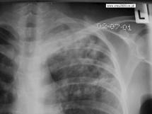
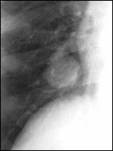
Subacute disseminated tuberculosis of lungs at incomplete absorbtion of foci and insufficient treatment passes to the chronic form. Sometimes from the beginning this form of tuberculosis runs across as a chronic disease. Roentgenological at chronic disseminated tuberculosis find out the foci of different closeness and size, which are located asymmetric among fibrous and emphysematous changed parenchym of lungs, cavities of disintegration, deformed and displaced roots of lungs. Focal tuberculosis more often develops as a result of acuteening of inflammatory process round a primary tubercular complex. In 1st, 2nd, 6th segments focal shades of different sizes, rounded form, small intensity appears on a background the increases lung pattern, with unclear contours; at the focal level noticeable pleura stratifications. Specific treatment results in absorbing of such foci during 2-3 months. At insufficient treatment and subsequent motion of disease next to the described soft "exudative" foci dense "productive" foci are determined by the sizes 4-6mm, middle intensity with clear contours, a lung pattern is rough, fibrous. Productive foci do not resolve under act of treatment, but compressing, diminish and calcificating. Infiltrative tuberculosis (see fig 17.4)develops as the perifocal inflammation round primary foci, as a result they meet between its, forming tubercular infiltrates of different sizes. Infiltrativne tuberculosis meets in 40-60% patients in which tuberculosis was finding first. a Fig.17.4 Infiltrative tuberculosis, tubercular cavity Clinical development of the disease reminds acute respiratory virial infections or pneumonia: acute beginning, cough, pain in breasts, and increase of the body temperature to 38-39 ° С. Roentgenological in 1st, 2nd, 6th segments infiltrates of different types are find out. Infiltrate is limited (clavicle) as round shade of small intensity, heterogeneous structure, wrong form, with unclear contours, by a size of 2-3 sm, disposed at the level of clavicle or under it. Nebulous infiltrate gives the shade small intensity, unclear outlined, which occupies all of segment, or his part. Lobar infiltrate which engulfs all lobe and tubercular is called lobitis. Infiltrate which is localized along contour of lungs lobe after motion of interlobar fissura, tubercular periscis-suritis is called. For all infiltrative processes characteristic a path of lymphangoitis towards the extended lung’s roots. Under act of treatment tubercular infiltrate is resolves, compressing with formation in them place of fibrous-cirrhotic changes and calcifications. At unfavorable course there is destruction in infiltrates, he tests caseous disintegration, by investigation formation of cavity can be and bronchogenous dissemination. Caseous pneumonia is the heaviest form of lungs tuberculosis, which is characterized by infiltration of all lobes and mushroom growth of destruction. The contours of infiltrative shade and focus of destruction are unequal and unclear. There is a rapid bronchogenic distribution of process and appearance of numerous segmental foci, feel like confluence and disintegration. Tuberculoma is the focus of caseous necrosis of the rounded form, which is marked off from surrounding pulmonary tissue a fibred connecting shell. Clinically tuberculoma often does not find out itself, however always positive reaction with tuberculin. Roentgenologicaly tuberkuloma has the appearance of the rounded intensive shade with clear, sometimes with unequal contours, heterogeneous structure, with sizes usually 2-4sm. Tuberculoma more often under a pleura in 1st 2nd 6th segments, that stipulates a presence nearby tuberculoma of pleuropulmonar scars, reduction of costal and interlobaroy pleura. In adjoining or in remote areas lungs are often observed different size the calcinated foci and fibrous changes.
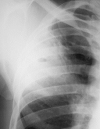
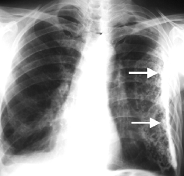
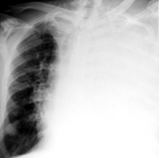
Fig.17.5 Tuberculoma In the phase of acuteening the external contours of tuberculoma become unclear as the result of inflammation; focuss appear in surrounding departments of lungs, pleurisy, and path of lymphangoitis. Cavernous and fibrous-cavernous tuberculosis arises out of any forms of tuberculose as the result of ill-timed exposure of disease, lately begun treatment, features of reactivity of organism and other, which results in progress of process and origin of caseous-nekrotic and destructive changes of pulmonary tissue. Cavernous tuberculosis characterizes a presence in lungs of stable cavity, by absence of perifocal inflammation and fibrotic changes in surrounding pulmonary tissue. A cavity contains caseous the masses, the internal surface of its thick wall is unequal. Roentgenological ring-shade of different size (more frequent 2-4sm) appears with external unclear and internal clear bay-shaped contours. On a background tubercular focus exposure cavities and areas of pneumosclerosis computer tomography is instrumental. With tearing away of caseous masses through a drening bronchus, the thickness of cavity`s wall diminishes, and an internal surface is evened. In the case of protracted motion a disease passes to the fibrous-cavernous form. Fibrous-cavernous tuberculosis is an active destructive form of tuberculosis which is characterized by chronic motion with the change of periods of acuting and calming down of process. Rentegnomorphologicaly this form of tuberculosis characterizes a presence in lungs of one or a few cavities with a thick thre-layer wall (internal caseous, middle granulation, external fibrous layers), and also development of sclerotic changes in adjoining pulmonary tissue, foci of sifting out of different remoteness, pleura reductions and lungs emphysema. The lung pattern of the staggered lobe is increased and deformed, root lungs on the staggered side deformed, displaced in anterior alion of sclerotic of the changed lobe, quite often extended. A pleura is unevenly thickened, pleura recesss fully or partly oblitereted. Such roentgenological pattern can remain permanent during many months and even years. Daughter's cavities appear in the case of progress of process, an inflammatory process spreads on the new areas which can engulf a lobe or all lung. Under act of treatment a cavity heals over and develops cirrhotic tuberculosis. Cirrhotic tuberculosis (see fig 17.6) is an eventual form of tuberculosis, for which characteristic are surplus excrescences of connecting tissue in lungs, that change alveolar parenchyma. Cirrhosis is always accompanied wrinkling and diminishing of lungs volume, or its lobes. Caseous foci, remaining cavities which can entail acuteening of process, are however saved. More often all cirrhotic changes are localized in superior lobes. Such roentgenological pattern appears at the cirrhosis of superior lobes: a lobe is considerably diminished in a volume, a root compressed, lifted up to the top, lung pattern is deformed, the areas of emphysema of pulmonary tissue appear, a parietal pleura is thickened, the organs of mediastinum are displaced toward a defeat, intercostal intervals are narrowed. A tubercular pleurisy (see fig 17.7) it is very often a complication of other forms of tuberculosis, however can be an independent form. Has those roentgenological signs, that and pleurisy of untubercular etiology.
Fig.17.6 Cirrhotic tuberculosis
Fig.17.7 Tubercular pleurisy
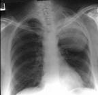
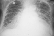
Extra-pulmonary tuberculosis Tuberculosis of superior respiratory tracts, trachea and bronches arises up as the result of the complicated motion of other clinical forms of lungs tuberculosis. Rqentgenological sign of tubercular damage of respiratory tracts appear mainly by computer tomography and bronchography. It is constriction of the large bronches lumen of different degree, sometimes their obturation. The walls of bronches are unevenly thickened, their internal contour is unequal. Coniotuberculosisis a tuberculosis of respiratory organs, combined with the dust professional diseases of lungs. More often tuberculosis is combined with silicosis (silicotuberculosis). A roentgenological pattern consists of aggregate of signs, characteristic for both diseases. Tuberculosis of interthoracal lymphatic nodes (tubercular bronchoadenitis) (see fig 17.8) is also the primary form of tuberculosis, which develops in 35-45% childrens and 1-3% adults, patients with tuberculosis. Patomorphological changes arise up mainly in bronchopulmonary lymphatic nodes of superior lobes of lungs and right lung`s middle lobe. a Fig.17.8 Tuberculosis of interthoracal lymphatic nodes, infiltrative and tumular forms Distinguish the infiltrative and tumular forms of tuberculosis of interthoracal lymphatic nodes. The infiltrative form of bronchoadenitis is observed more often than tumular; it arises up as the result of passing of process to the walls of bronches and roentgenological characterized increasing shade of root lungs, which has homogeneous character and unclear contours. The tumular form of tubercular bronchoadenitis characterizes the greater sizes of lymphatic nodes in composition a root, which has a clear external contour, but not differentiated separately. These forms in the case of dynamic supervision can pass to each other. Increase of bronhopulmonary and tracheobronchialy lymphatic nodes better appears by computer tomography. For the disseminated form of tuberculosis of lungs a characteristic presence in lungs of the plural dissipated tubercular foci is as a result of his distribution by lympho-, hemato- and bronchogenic way. After motion disseminated lungs tuberculosis distinguish acute, subacute and chronic. For all the forms of disseminated tuberculosis of lungs is characteristic bilateral symmetric defeat. Miliar tuberculosis (see fig 17.9) is shallow focuss 2 – 3mm thickly will strike internal organs a diameter including lungs which predetermines heavy clinical motion as the result of defeat of considerable volumes of tissues. а Fig.17.9 Lungs miliar tuberculosis
Radio diagnostics of the breathing organs tumours|swelling|. Radio diagnostics of the urgent states at the breathing organs pathology.|composition| Algorithm of the breathing organs radioexamination.
The radio diagnostics of the urgent states at pathology of breathing organs. Roentgenologic, radionuclear|, ultrasonic, magnetically-resonance imaging signs of the urgent states – lungs edema, hydrothorax|, pneumothorax|, acute pulmonary thromboembolism|, foreign bodies in bronches, traumatic damages. Choice of the radio examination method for diagnostics of the concrete urgent state. Algorithm of the breathing organs radio examination (fluorography |, sciagraphy, fluoroscopy |, linear tomography |, computer tomography |, magnetically-resonance imaging|, scintigraphy |, SPECТ|, PAT, radionuclear | scanning, radiometry, and sonography |). Date: 2014-12-28; view: 2431
|
 б
б 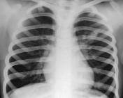 в
в 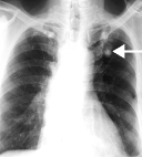
 b
b 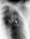
 b
b 
 б
б