CATEGORIES:
BiologyChemistryConstructionCultureEcologyEconomyElectronicsFinanceGeographyHistoryInformaticsLawMathematicsMechanicsMedicineOtherPedagogyPhilosophyPhysicsPolicyPsychologySociologySportTourism
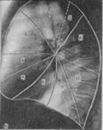
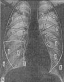
The thoracic organs roentgenanatomyOn survey X-ray film (see fig 15.6) in the anterior view the 7 pair of ribs are visualized. Lower ribs are partly hidden by the diaphragmal shadow and organs of abdominal region. Cartilaginous parts of the ribs is invisible on a X-radiographies (sciagrams, X-ray). After 30 years calcination arises in costal cartilage of the 1th ribs. A shoulder-blade and clavicle is well noticeable at fluoroscopy and on the sciagram. At the patient correct position in the anterior view, the clavicle sternumends are disposed symmetric on identical distance from a vertebral column.The sternum in the anterior view is invisible. In a lateral projection the shade of sternum forms a contour of chest.
Fig.15.6 Sciagram of the thoracal|mew| organs (marked elements of anatomic formations|formations|):1-trachea; 2- cupulaes of diaphragm; 3-right and left main|head,leading| bronches; 4-arc of the right auricle; 5-descendence| part|portion| of aorta; 6-aortal arc; 7-projection of subclavial arteries; 8-left heart border|line,boundary| (from above to downof the| arc: ascending part|portion| of aorta, the cone of pulmonary artery, auricle of the left auricle and left ventricle); 9-azygos vein; 10-horizontal interlobar| fissura; 11-intermediate pulmonary artery; 12-left main|head,leading| pulmonary artery; 13-lungs roots; 14-segmentar arteries(sub-root| area|zone|); 15-vessels of a 2nd intervertebral| interval|space| (in the norm|standard| 3 mm| in a diameter); 18-back bends of pleura;19-clavicules; Diaphragm limits the thorax from the organs of abdominal region. Diaphragm together with the organs of abdominal region has intensive shade. Diaphragm consists from left and right cupulaes (domes). Under left cupula of diaphragm is radiolucent zone of air-filled gastric funduss. The right cupula of diaphragm takes a place at the level of anterior ends of 5-6th ribs, and left - on one rib below. The costodiaphragmatic recess are noticeable in the lateral corners of cupulas of diaphragm, and in medial corners are costomediastinal recesses. In lateral projection anterior costodiaphragmatic recesses are placed higher then posterior costodiaphragmatic recess. In the anterior view are defined such pulmonary fields: 1) apex of lung – there are areas of lungs above clavicles; 2) superior lung fields - there are areas of lungs between clavicles and anterior parts of the 2-nd ribs; 3) middle lung fields - there are areas of lungs between anterior parts of 2-nd and 4-th ribs; 4) inferior lung fields - there are areas of lungs between anterior parts of 4 rib and diaphragm. In a lateral projection well is evidently the shade of sternum, blade-bone, from 6th to 9th thorasic vertebraes and shades of ribs. Narrower and more expressly are shades from the ribs near the X-ray film. Radiolucent area between a sternum and a heart is called retrosternal space, radiolucent area between the heart and vertebral column is called retrocardial space. Shadow of lungs root are formed from pulmonary arteries, veins, bronches, lymphatic vessels and nodes. In the anterior view this shade is not homogeneous structure, located at the level of anterior ends from 2 to 4-th ribs, in a width about 2 cm, occupies the not more than quarter of transversal size of lung. A root of left lung places higher than right, and his lower part closes by cardiac shade. In a lateral projection roots of lungs are under the aortal arch. Most thoracic lymphatic nodes (see fig 15.7) are in the lungs roots and mediastinum. In the case of inflammatory and tumour processes they are increased and shade of root becomes homogeneous and unstructured.
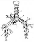
Fig.15.7 Intrathorasic lymphatic nodes 1– paratracheal; 2 – tracheobronchial; 3 – bifurcational; 4 – bronchopulmonary
Linear shades of pulmonary vessels which comes away from the roots of lungs to lungs parenchima are called lung pattern, interlacing, are formed ansiform structure. The caliber of these shades gradually diminishes in anterior alion from roots and on 2 sm does not reach to the thorasic wall. Parietal pleura can be traced in the anterior view as the thin arched strip which stretches from the apex of lateral surface lungs. Interlobar pleura and clear border between pulmonary lobes in the anterior view is not traced. The anterior view of oblique fissura goes from the 3rd thorasic vertebra obliquely downward to the lateral surface of 4th rib (at the posterior surface of a body) and farther obliquely downward to the diaphragm. A horizontal interlobar fissura (is in right lung) goes horizontally from the lateral surface of the 4th rib to lungs root. In a lateral projection the pleura of oblique interlobar fissura forms thin ("hairy") shade, that going from the level of 4th thorasic vertebra obliquely downward and anterior to the diaphragm. A horizontal fissura in a lateral projection begins from oblique at the level of root lungs and goes ahead to the thorasic wall horizontally (see fig 15.8).
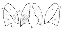
Fig.15.8 projection of lobes and interlobar fissuras is on a sciagram (chart) a) right lateral projection; b) anterior view; c) left lateral projection. 1-superior lobes; 2-middle lobe; 3-inferior lobes; 4-horizontal fissura; 5-oblique fissura Left lung consists from two lobes, right lung consists from three lobes. Lobes consist from segments, which have a wrong pyramids form. The apexes of segments are dicected to the root, the bases of segments - to the surface lungs (see fig 15.9 and Table 15.1).
Roentgenological border between segments of lung are not present; however for the defining localization of pathological process it is necessary to know the topographical structure of bronchial tree, lobes and segments. A right lung consists of 3 lobes 10 segments, left - from 2 lobes and 10 segments. a Fig.15.9 The schematic image of the lungs segments. Anterior view (a), right lateral (b), left lateral (c) projections 1 - apex, 2 - back, 3 - anterior , 4 - lateral (in left lungs - superior uvular), 5 - medial (in left lungs - lower uvular), 6 – superior lower lobe; 7 - cardial (in left lungs absents often), 8 – anterior bazal, 9 – lateral basal, 10 - back basal. Table 15.1 Lungs segments
Date: 2014-12-28; view: 1648
|
 b
b 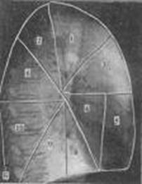 c
c